. 2017 Jul 3;216(7):1949-1957.
doi: 10.1083/jcb.201610113. Epub 2017 May 31.

Persistent mTORC1 signaling in cell senescence results from defects in amino acid and growth factor sensing

Affiliations

Persistent mTORC1 signaling in cell senescence results from defects in amino acid and growth factor sensing

Bernadette Carroll et al. J Cell Biol. .

Abstract

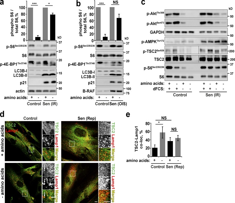
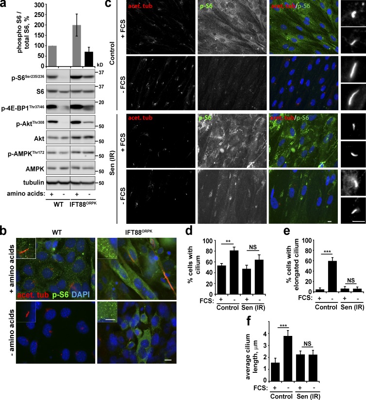
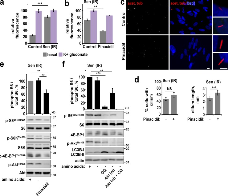
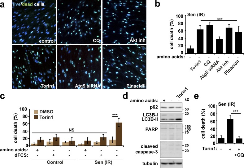
Mammalian target of rapamycin complex 1 (mTORC1) and cell senescence are intimately linked to each other and to organismal aging. Inhibition of mTORC1 is the best-known intervention to extend lifespan, and recent evidence suggests that clearance of senescent cells can also improve health and lifespan. Enhanced mTORC1 activity drives characteristic phenotypes of senescence, although the underlying mechanisms responsible for increased activity are not well understood. We have identified that in human fibroblasts rendered senescent by stress, replicative exhaustion, or oncogene activation, mTORC1 is constitutively active and resistant to serum and amino acid starvation. This is driven in part by depolarization of senescent cell plasma membrane, which leads to primary cilia defects and a resultant failure to inhibit growth factor signaling. Further, increased autophagy and high levels of intracellular amino acids may act to support mTORC1 activity in starvation conditions. Interventions to correct these phenotypes restore sensitivity to the mTORC1 signaling pathway and cause death, indicating that persistent signaling supports senescent cell survival.


Figures

Figure 1.
Figure 1.
PI3K/Akt/mTOR/autophagy are insensitive to starvation in senescent cells. (a) Immunoblot analysis of mTORC1 and autophagy activity in control and senescent (Sen(IR); 30 d after 20 Gy x-ray irradiation) primary human fibroblasts after overnight serum starvation with or without 1-h amino acid starvation. (b) As in panel a, but senescence was achieved by transduction of cells with oncogenic mutant B-RAFV600E (oncogene-induced senescence, OIS). (c) Immunoblot analysis of signaling cascades upstream of mTORC1 after starvation protocols as described in panel a. (d and e) Proliferating or replicative senescent (Sen(Rep)) cells were treated as in a, fixed, and immunostained for antibodies against TSC2 and Lamp1. White arrows indicate recruitment of TSC2 to lysosomes (d). Colocalization between Lamp1 and TSC2 (e) was analyzed. Error bars represent SEM; all experiments, n = 3 (for colocalization, at least 10 cells imaged per experimental repeat). Student’s t test performed between groups: *, P < 0.05; ***, P < 0.001; NS, not significant. Bars, 10 µm.
Figure 2.
Figure 2.
Senescent cells fail to induce primary cilium elongation. (a and b) Wild-type and IFT88ORPK chondrocytes (which are deficient in primary cilia; Wann et al., 2012) were grown until confluent and subjected to serum starvation overnight. Cells were either lysed and subjected to Western blotting (a) or fixed and immunostained with antibodies against acetylated tubulin and phospho-S6 (b). (c–f) Confluent control and senescent (20 Gy irradiation; Sen (IR)) fibroblasts were serum-starved overnight (–FCS), fixed, and immunostained for acetylated tubulin and phospho-S6 (c). Percentage of cells with primary cilium (d), percentage of cells with elongated primary cilium (e), and mean cilium length (f) were quantified. Error bars represent SEM; all experiments, n = 3 (for immunofluorescence, at least five fields of view imaged per experimental repeat). Student’s t test performed between groups: **, P < 0.01; ***, P < 0.001; NS, not significant. Bars: (main) 10 µm; (insets) 5 µm.
Figure 3.
Figure 3.
Restoration of membrane potential in senescent cells promotes cilia elongation. (a) Resting and maximum membrane potential (maximum achieved by incubation for 5 min with 80 mM potassium gluconate) were measured in control and senescent (20 Gy irradiation; Sen (IR)) fibroblasts using DiSBAC2(3) dye. Depolarization is indicated by increased fluorescence. Data are represented relative to maximum. (b) Senescent cells treated as in a but in the presence or absence of 100 µM pinacidil overnight. (c and d) Senescent fibroblasts were treated with pinacidil overnight in serum-free medium. Cells were fixed and stained for acetylated tubulin (c). Percentage of cells with cilia and cilia length were quantified (d). (e) Senescent fibroblasts were starved of serum overnight and amino acids for 1 h in the presence or absence of pinacidil as indicated, lysed, and analyzed by immunoblot for mTORC1 activity. (f) Senescent fibroblasts were starved of serum overnight and amino acids for 1 h in the presence or absence of 50 µM autophagy inhibitor chloroquine (CQ), 10 µM Akt inhibitor, or both inhibitors as indicated. Cells were lysed and immunoblotted. Error bars represent SEM; all experiments, n = 3 (for immunofluorescence, at least five fields of view imaged per experimental repeat). Student’s t test performed between groups: **, P < 0.01; ***, P < 0.001; NS, not significant. Bars: (main) 10 µm; (insets) 5 µm.
Figure 4.
Figure 4.
Persistent Akt/mTORC1 signaling supports senescent cell survival and can be exploited to specifically promote cell death. (a and b) Senescent fibroblasts were starved of serum and amino acids overnight in the presence or absence of the mTOR kinase inhibitor 200 nM Torin1 (overnight), 50 µM autophagy inhibitor chloroquine (CQ; 4 h), or 10 µM Akt inhibitor (4 h) as indicated. Cells were incubated with fluorescent ReadyProbe Cell Viability reagents (a), and percentage cell death was quantified (b). (c) Control or senescent (20 Gy irradiation; Sen(IR)) fibroblasts were starved of serum and amino acids in the presence or absence of 200 nM Torin1 for 24 h. Cells were incubated with fluorescent ReadyProbe Cell Viability reagents, and cell viability was quantified (c). (d) Senescent fibroblasts were starved in the presence or absence of Torin1. Cells were lysed and analyzed for markers of apoptosis (PARP and cleaved caspase-3) and autophagy (p62 and LC3). (e) Senescent fibroblasts were starved in the presence or absence of Torin1 and chloroquine as indicated. Percentage cell death was analyzed by ReadyProbe cell viability reagents. Error bars represent SEM; for all experiments, n = 3 (for immunofluorescence, at least five fields of view imaged per experimental repeat). Student’s t test performed between groups: ***, P < 0.001; NS, not significant. Bar, 30 µm.

References

    1. Baker D.J., Wijshake T., Tchkonia T., LeBrasseur N.K., Childs B.G., van de Sluis B., Kirkland J.L., and van Deursen J.M.. 2011. Clearance of p16Ink4a-positive senescent cells delays ageing-associated disorders. Nature. 479:232–236. 10.1038/nature10600 - DOI - PMC - PubMed
    1. Baker D.J., Childs B.G., Durik M., Wijers M.E., Sieben C.J., Zhong J., Saltness R.A., Jeganathan K.B., Verzosa G.C., Pezeshki A., et al. . 2016. Naturally occurring p16(Ink4a)-positive cells shorten healthy lifespan. Nature. 530:184–189. 10.1038/nature16932 - DOI - PMC - PubMed
    1. Bishop C.L., Bergin A.M., Fessart D., Borgdorff V., Hatzimasoura E., Garbe J.C., Stampfer M.R., Koh J., and Beach D.H.. 2010. Primary cilium-dependent and -independent Hedgehog signaling inhibits p16(INK4A). Mol. Cell. 40:533–547. 10.1016/j.molcel.2010.10.027 - DOI - PubMed
    1. Boehlke C., Kotsis F., Patel V., Braeg S., Voelker H., Bredt S., Beyer T., Janusch H., Hamann C., Gödel M., et al. . 2010. Primary cilia regulate mTORC1 activity and cell size through Lkb1. Nat. Cell Biol. 12:1115–1122. 10.1038/ncb2117 - DOI - PMC - PubMed
    1. Breslin L., Prosser S.L., Cuffe S., and Morrison C.G.. 2014. Ciliary abnormalities in senescent human fibroblasts impair proliferative capacity. Cell Cycle. 13:2773–2779. 10.4161/15384101.2015.945868 - DOI - PMC - PubMed

MeSH terms

Substances