. 2017 Jul 27;91(16):e00519-17.
doi: 10.1128/JVI.00519-17. Print 2017 Aug 15.

Discovery and Mechanistic Study of Benzamide Derivatives That Modulate Hepatitis B Virus Capsid Assembly

Affiliations

Discovery and Mechanistic Study of Benzamide Derivatives That Modulate Hepatitis B Virus Capsid Assembly

Shuo Wu et al. J Virol. .

Abstract

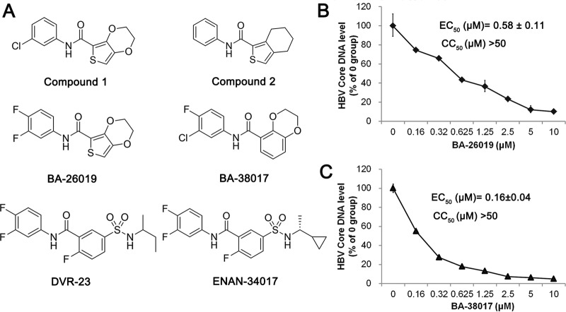
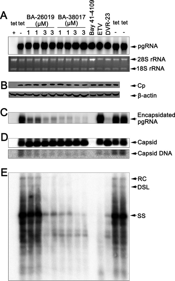
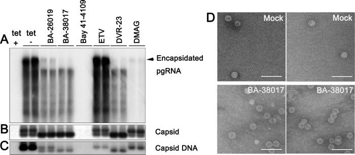
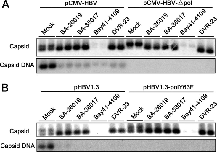
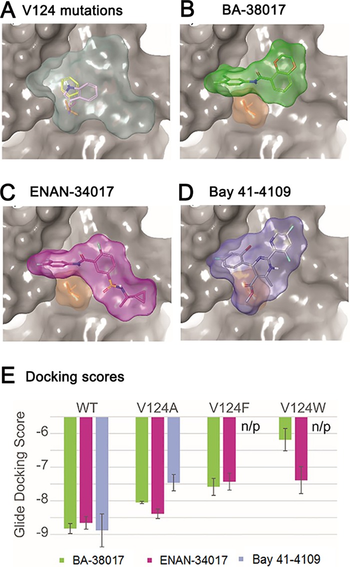
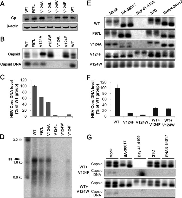
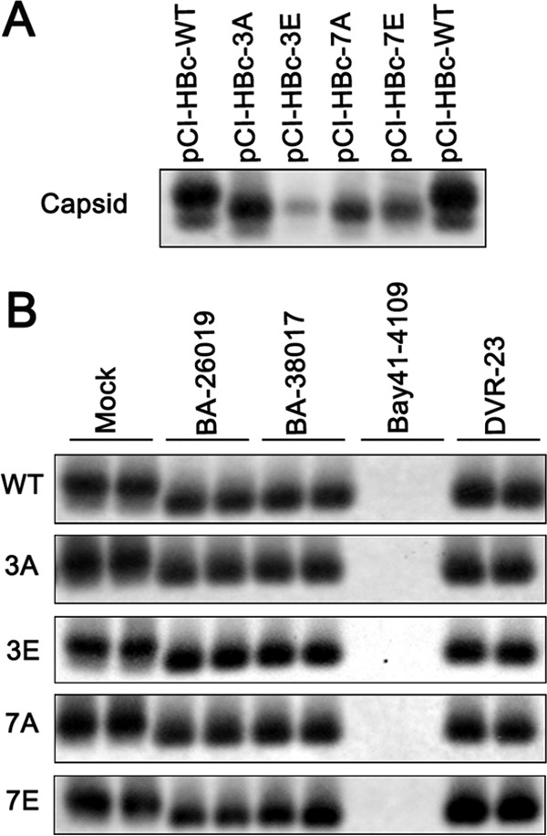
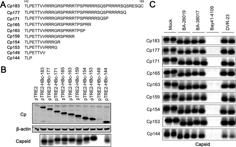
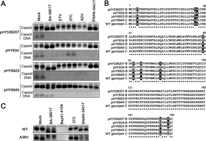
Chronic hepatitis B virus (HBV) infection is a global public health problem. Although the currently approved medications can reliably reduce the viral load and prevent the progression of liver diseases, they fail to cure the viral infection. In an effort toward discovery of novel antiviral agents against HBV, a group of benzamide (BA) derivatives that significantly reduced the amount of cytoplasmic HBV DNA were discovered. The initial lead optimization efforts identified two BA derivatives with improved antiviral activity for further mechanistic studies. Interestingly, similar to our previously reported sulfamoylbenzamides (SBAs), the BAs promote the formation of empty capsids through specific interaction with HBV core protein but not other viral and host cellular components. Genetic evidence suggested that both SBAs and BAs inhibited HBV nucleocapsid assembly by binding to the heteroaryldihydropyrimidine (HAP) pocket between core protein dimer-dimer interfaces. However, unlike SBAs, BA compounds uniquely induced the formation of empty capsids that migrated more slowly in native agarose gel electrophoresis from A36V mutant than from the wild-type core protein. Moreover, we showed that the assembly of chimeric capsids from wild-type and drug-resistant core proteins was susceptible to multiple capsid assembly modulators. Hence, HBV core protein is a dominant antiviral target that may suppress the selection of drug-resistant viruses during core protein-targeting antiviral therapy. Our studies thus indicate that BAs are a chemically and mechanistically unique type of HBV capsid assembly modulators and warranted for further development as antiviral agents against HBV.IMPORTANCE HBV core protein plays essential roles in many steps of the viral replication cycle. In addition to packaging viral pregenomic RNA (pgRNA) and DNA polymerase complex into nucleocapsids for reverse transcriptional DNA replication to take place, the core protein dimers, existing in several different quaternary structures in infected hepatocytes, participate in and regulate HBV virion assembly, capsid uncoating, and covalently closed circular DNA (cccDNA) formation. It is anticipated that small molecular core protein assembly modulators may disrupt one or multiple steps of HBV replication, depending on their interaction with the distinct quaternary structures of core protein. The discovery of novel core protein-targeting antivirals, such as benzamide derivatives reported here, and investigation of their antiviral mechanism may lead to the identification of antiviral therapeutics for the cure of chronic hepatitis B.

Keywords: antiviral agents; capsid assembly; hepatitis B virus.


Figures

FIG 1
FIG 1
Identification of benzamide derivatives (BAs) that inhibit HBV replication. (A) Chemical structures of two primary screening “hits,” two optimized lead benzamide derivatives, BA-26019 and BA-38017, as well as two reference sulfamoylbenzamide derivatives (DVR-23 and ENAN-34017) used in this study are presented. (B and C) AML12HBV10 cells were treated with the indicated concentrations of compound BA-26019 or BA-38017 for 2 days. Cytoplasmic HBV core DNAs were extracted and quantified by a qPCR assay and expressed as the percentage of the mock-treated controls. The means and standard deviations (n = 4) were plotted. The cytotoxicity was determined by an MTT assay.
FIG 2
FIG 2
Antiviral mechanism of BA derivatives against HBV. AML12HBV10 cells were cultured in the presence of tetracycline (tet +) or in the absence of tetracycline and mock treated (tet −) or treated with the indicated concentrations of the compound BA-26019 (1 and 3 μM), BA-38017 (1 and 3 μM), ETV (1 μM), DVR-23 (2 μM), or Bay 41-4109 (2 μM) for 2 days. (A) Intracellular viral RNA was measured by Northern blotting hybridization. 28S and 18S rRNA were used as loading controls. (B) HBV core protein (Cp) expression was detected by Western blotting with a rabbit polyclonal antibody (Dako). β-Actin served as the loading control. The results were derived from two separate gels, as indicated. (C) Encapsidated pgRNA was determined by Northern blotting. (D) The total amounts of capsids were determined by a particle gel assay in a 1.0% agarose gel electrophoresis. Capsid-associated HBV DNA was detected by hybridization upon alkaline treatment of nucleocapsids on the membrane following the particle gel assay. (E) HBV DNA replication intermediates were extracted and determined by Southern blotting. A 32P-labeled full-length minus-strand-specific riboprobe was used for Southern blot analysis. RC, relaxed circular DNA; DSL, double-stranded linear DNA; SS, single-strand, negative-polarity DNA.
FIG 3
FIG 3
BA and SBA compounds alter HBV capsid migration in native agarose gel electrophoresis. AML12HBV10 cells were cultured in the presence of tetracycline (tet +) or in the absence of tetracycline and mock treated (tet −) or treated with BA-26019 (5 μM), BA-38017 (5 μM), Bay 41-4109 (2 μM), ETV (1 μM), DVR-23 (2 μM), or DMAG (0.15 μM) for 2 days. (A) Encapsidated pgRNA was extracted and detected by Northern blotting hybridization. (B) The capsids were separated in a 1.5% agarose gel electrophoresis, transferred onto a nylon membrane, and detected by a rabbit polyclonal antibody against core protein (Dako). (C) Capsid-associated HBV DNA was detected by hybridization with a 32P-labeled full-length HBV minus-strand-specific riboprobe upon alkaline treatment of the membrane following the particle gel assay. (D) Two representative electron micrographs of capsids purified from AML12HBV10 cells mock treated (upper panel) or treated with 5 μM BA-38017 (lower panel) for 2 days. Bar, 100 nm.
FIG 4
FIG 4
BA derivatives alter the capsid assembly in a viral DNA polymerase-independent manner. HepG2 cells were transiently transfected with plasmid pCMV-HBV or pCMV-HBV-Δpol (A) or with pHBV1.3 or pHBV1.3polY63F (B). Six hours posttransfection, the cells were mock treated or treated with 5 μM BA-26019, 5 μM BA-38017, 2 μM Bay 41-4109, or 2 μM DVR-23 for 48 h. The capsids were separated by 1.5% agarose gel electrophoresis, transferred onto a nylon membrane, and detected by a rabbit polyclonal antibody against HBV core protein (Dako). Capsid-associated HBV DNA was detected by hybridization with a 32P-labeled full-length HBV minus-strand-specific riboprobe upon alkaline treatment of the membrane following the particle gel separation.
FIG 5
FIG 5
Structural simulation and docking analysis. (A) Binding site (light blue area) between the core protein dimer-dimer interface, with A, F, and W mutations of V124 shown as sticks. All the compounds bound at the HAP pocket. (B) Binding pose of 38017, green compound; (C) binding pose of 34017, magenta compound; (D) binding pose of Bay 41-4109, gray compound. V124 is shown in tan for each pose, and part of chain C was hidden to visualize the binding site. (E) Docking energies for each of the compounds with each mutant. n/p, no pose reported during docking.
FIG 6
FIG 6
Replacement of core protein valine 124 with amino acids with different sizes of hydrophobic side chains confers resistance to BA and SBA compound-induced alteration of capsid assembly. HepG2 cells were transfected with plasmid pHBV1.3 or pHBV1.3-derived plasmids expressing core protein with F97L, V124A, V124L, V124F, or V124W mutation. The cells were harvested at 48 h posttransfection. (A) HBV core protein (Cp) expression was detected by Western blotting with a rabbit polyclonal antibody (Dako). (B) Capsids and capsid-associated HBV DNA were detected by a particle gel assay. (C) Capsid-associated HBV DNAs in the transfected cells were quantified by a qPCR assay, and the results were plotted as the percentage of the amounts in pHBV1.3-transfected cells (n = 4). (D) HBV DNA replication intermediates were detected by Southern blotting. (E) HepG2 cells were transfected with plasmid pHBV1.3 or pHBV1.3-derived plasmids expressing core protein with F97L, V124A, V124L, V124F, or V124W. Six hours posttransfection, the cells were mock treated or treated with 5 μM BA-38017, 10 μM 3TC, 2 μM Bay 41-4109, or 5 μM ENAN-34017 for 48 h. Cytoplasmic capsids were determined by a 1.5% agarose particle gel assay. (F) Capsid-associated HBV DNAs in the transfected cells were quantified by a qPCR assay, and the results were plotted as the percentage of the amounts in pHBV1.3-transfected cells (n = 4). (G) HepG2 cells were cotransfected with pHBV1.3 and pHBV1.3coreV124F or pHBV1.3coreV124W in a molar ratio of 1:1. Six hours posttransfection, the cells were mock treated or treated with BA-38017 (5 μM), 3TC (10 μM), Bay 41-4109 (2 μM), or ENAN-34017 (5 μM) for 72 h. Cytoplasmic capsids were analyzed by a particle gel assay. Capsid-associated HBV DNA was quantified by hybridization with a 32P-labeled full-length HBV minus-strand-specific riboprobe.
FIG 7
FIG 7
The phosphorylation status of the CTD does not affect BA and SBA modulation of capsid assembly. (A) AML12 cells were transfected with plasmid pCI-HBc, pCI-HBc-3A, pCI-HBc-3E, pCI-HBc-7A, or pCI-HBc-7E. The cells were harvested at 48 h posttransfection. The cytoplasmic capsids were analyzed by a particle gel assay. (B) AML12 cells were transfected with the indicated core protein-expressing plasmid. Six hours posttransfection, the cells were left untreated or treated with 5 μM BA-26019, 5 μM BA-38017, 2 μM Bay 41-4109, or 2 μM DVR-23 for 48 h. The cytoplasmic capsids were analyzed by a particle gel assay.
FIG 8
FIG 8
The arginine-rich CTD of HBV core protein does not play a role in BA and SBA modulation of capsid assembly. (A) Sequences of wild-type or C-terminally truncated HBV core proteins expressed by plasmids. (B) AML12 cells were transfected with vector plasmid or a plasmid expressing either wild-type full-length core protein or the indicated C-terminally truncated core protein. The cells were harvested at 48 h posttransfection. Core protein (Cp) expression was detected by Western blotting (top), and β-actin served as the loading control (middle). The cytoplasmic capsids were analyzed by a particle gel assay (bottom). (C) AML12 cells were transfected with a plasmid expressing either wild-type core protein or the indicated C-terminally truncated core protein. Six hours posttransfection, the cells were left untreated (NT) or treated with 5 μM 26019, 5 μM BA-38017, 2 μM Bay 41-4109, or 2 μM DVR-23 for 48 h. The cytoplasmic capsids were analyzed by a particle gel assay.
FIG 9
FIG 9
Effects of BA and SBA compounds on capsid assembly and DNA replication of clinically isolated strains of HBV. (A) HepG2 cells were transfected with the indicated plasmid containing a replication-competent HBV genome derived from Chinese patients. Six hours posttransfection, the cells were mock treated or treated with BA-38017 (5 μM), ETV (0.125 μM), 3TC (1 μM), ADV (0.2 μM), or ENAN-34017 (5 μM) for 72 h. Cytoplasmic capsids were analyzed by a particle gel assay. Capsid-associated HBV DNA was quantified by hybridization with a 32P-labeled full-length HBV minus-strand-specific riboprobe upon alkaline treatment of the membrane following the particle gel assay. (B) Amino acid sequence alignment with highlights of mutations. (C) HepG2 cells were transfected with plasmid pTRE2-HBc or pTRE2-HBcA36V. The transfected cells were treated with BA-38017 (5 μM), Bay 41-4109 (2 μM), 3TC (1 μM), or ENAN-34017 (5 μM) for 72 h. Cytoplasmic capsids were analyzed by a particle gel assay.

References

    1. GBD 2013 Mortality and Causes of Death Collaborators. 2015. Global, regional, and national age-sex specific all-cause and cause-specific mortality for 240 causes of death, 1990-2013: a systematic analysis for the Global Burden of Disease Study 2013. Lancet 385:117–171. doi:10.1016/S0140-6736(14)61682-2. - DOI - PMC - PubMed
    1. Ott JJ, Stevens GA, Groeger J, Wiersma ST. 2012. Global epidemiology of hepatitis B virus infection: new estimates of age-specific HBsAg seroprevalence and endemicity. Vaccine 30:2212–2219. doi:10.1016/j.vaccine.2011.12.116. - DOI - PubMed
    1. Dienstag JL. 2009. Benefits and risks of nucleoside analog therapy for hepatitis B. Hepatology 49:S112–S121. doi:10.1002/hep.22920. - DOI - PubMed
    1. Perrillo R. 2009. Benefits and risks of interferon therapy for hepatitis B. Hepatology 49:S103–S111. doi:10.1002/hep.22956. - DOI - PubMed
    1. Block TM, Gish R, Guo H, Mehta A, Cuconati A, Thomas London W, Guo JT. 2013. Chronic hepatitis B: what should be the goal for new therapies? Antiviral Res 98:27–34. doi:10.1016/j.antiviral.2013.01.006. - DOI - PMC - PubMed

Publication types

MeSH terms