. 2017 May 31;9(392):eaaf6295.
doi: 10.1126/scitranslmed.aaf6295.

Complement C3 deficiency protects against neurodegeneration in aged plaque-rich APP/PS1 mice

Affiliations

Complement C3 deficiency protects against neurodegeneration in aged plaque-rich APP/PS1 mice

Qiaoqiao Shi et al. Sci Transl Med. .

Abstract

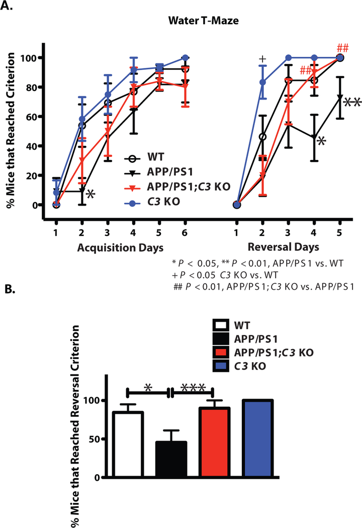
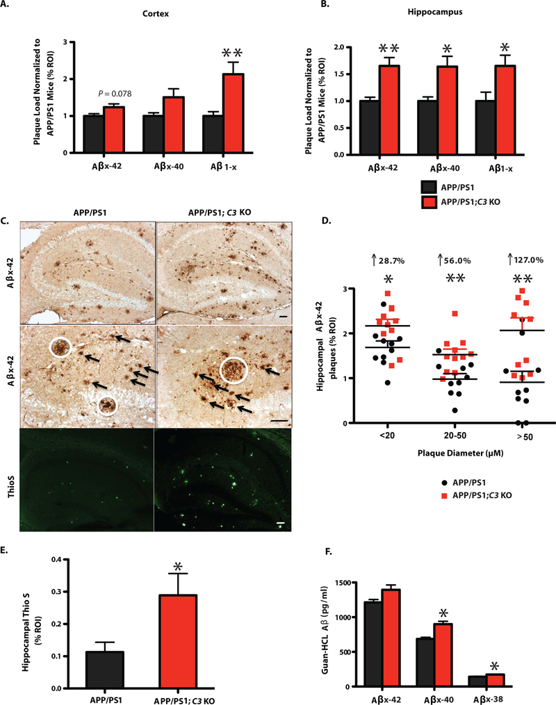
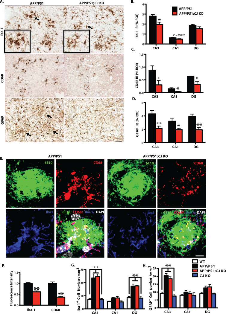
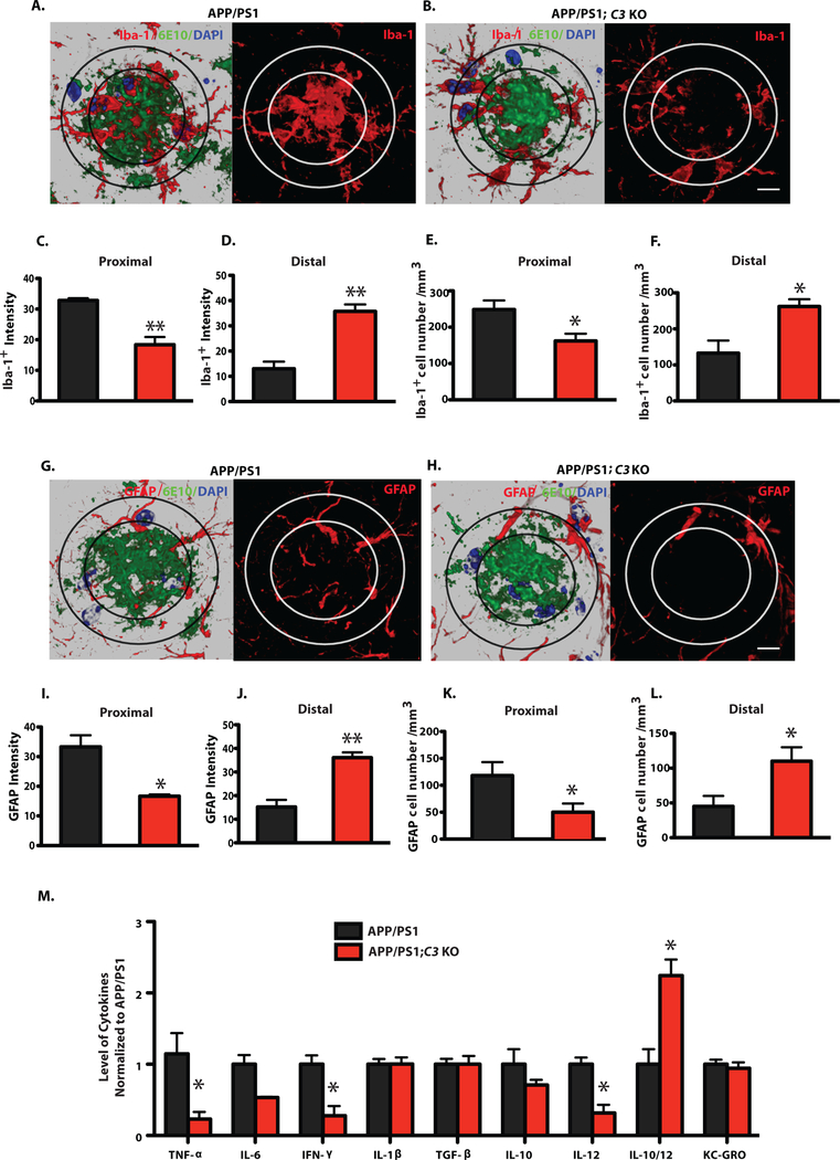
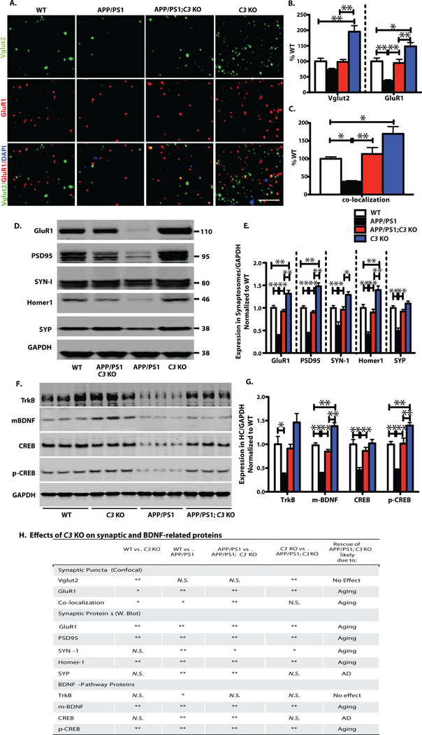
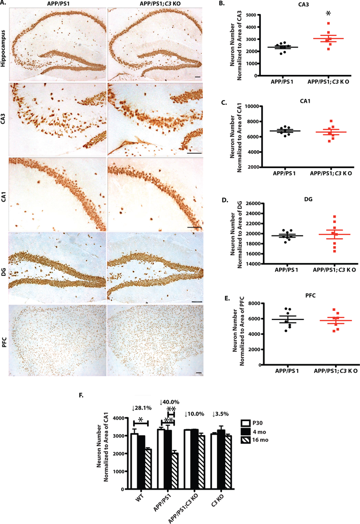
The complement cascade not only is an innate immune response that enables removal of pathogens but also plays an important role in microglia-mediated synaptic refinement during brain development. Complement C3 is elevated in Alzheimer's disease (AD), colocalizing with neuritic plaques, and appears to contribute to clearance of Aβ by microglia in the brain. Previously, we reported that C3-deficient C57BL/6 mice were protected against age-related and region-specific loss of hippocampal synapses and cognitive decline during normal aging. Furthermore, blocking complement and downstream iC3b/CR3 signaling rescued synapses from Aβ-induced loss in young AD mice before amyloid plaques had accumulated. We assessed the effects of C3 deficiency in aged, plaque-rich APPswe/PS1dE9 transgenic mice (APP/PS1;C3 KO). We examined the effects of C3 deficiency on cognition, Aβ plaque deposition, and plaque-related neuropathology at later AD stages in these mice. We found that 16-month-old APP/PS1;C3 KO mice performed better on a learning and memory task than did APP/PS1 mice, despite having more cerebral Aβ plaques. Aged APP/PS1;C3 KO mice also had fewer microglia and astrocytes localized within the center of hippocampal Aβ plaques compared to APP/PS1 mice. Several proinflammatory cytokines in the brain were reduced in APP/PS1;C3 KO mice, consistent with an altered microglial phenotype. C3 deficiency also protected APP/PS1 mice against age-dependent loss of synapses and neurons. Our study suggests that complement C3 or downstream complement activation fragments may play an important role in Aβ plaque pathology, glial responses to plaques, and neuronal dysfunction in the brains of APP/PS1 mice.


Figures

Fig. 1.
Fig. 1.
APP/PS1;C3 KO mice showed a significant improvement in cognitive flexibility (reversal) compared to APP/PS1 mice at 16 months of age. A. Percent of mice that reached criterion (≥ 80% correct choices on each individual day) in WTM test. Compared to WT mice, APP/PS1 mice were impaired in acquisition (Days 2) and reversal learning and memory (Days 4 and 5) (*p < 0.05; ** p < 0.01). APP/PS1;C3 KO mice performed significantly better than APP/PS1 mice (## p < 0.01), but similar to WT and C3 KO mice, in the reversal test on Days 4 and 5, suggesting better flexibility in APP/PS1;C3 KO mice compared to APP/PS1 mice. B. In total, fewer APP/PS1 mice reached the reversal criterion (≥80% correct choices over two consecutive days) (*p < 0.05), while the percent of WT, C3 KO and APP/PS1;C3 KO mice that reached criterion in the reversal test was significantly higher compared to APP/PS1 mice (***p < 0.001), indicating that C3-deficiency in APP/PS1 mice had both age-dependent and AD-related effects (WT, n=13; APP/PS1, n=11; APP/PS1;C3 KO, n=10; C3 KO, n = 11). Tests were assessed using one-way ANOVA followed by Fisher’s PLSD post hoc test.
Fig. 2.
Fig. 2.
Increased Aβ plaque deposition and Aβ levels by ELISA in 16 month-old APP/PS1;C3 KO mice. A,B. Quantification of Aβx-42, Aβx-40 and Aβ1-x (3D6) IR revealed increased plaque burden in cortex (A) and hippocampus (B) of 16-month-old APP/PS1;C3 KO mice compared to APP/PS1 mice. (* p < 0.05, ** p < 0.01 vs. APP/PS1 mice; n= 9 mice; 6 equidistant planes 150 μm apart; independent unpaired t-test per Aβ species). C. Aβx-42 IR and Thioflavin-S-positive plaque load was higher in the hippocampus of C3-deficient APP/PS1 mice vs. APP/PS1 mice. White circles indicate large plaques (> 50 μm); arrows indicate medium size plaques (> 20 but < 50 μm ). Scale bar = 50 μm. D. Quantification of small, medium and large hippocampal plaques confirmed an increased plaque load in APP/PS1;C3 KO mice, especially for large plaques (* p < 0.05, ** p < 0.01; n = 10; independent unpaired t-tests per plaque size category). E. Quantification of Thio-S in hippocampus confirmed an increase in fibrillar plaques in APP/PS1; C3 KO mice (* p < 0.05 vs APP/PS1 mice; n=6; 3 equidistant planes 300 μm apart; unpaired t-test). F. Increased insoluble cerebral Aβx-40 and Aβx-38 levels were found in APP/PS1;C3 KO mice compared with APP/PS1 (* p < 0.05; n=8) (independent unpaired t-tests per Aβ species).
Fig. 3.
Fig. 3.
Morphological changes associated with glial activation were reduced in 16-month-old APP/PS1;C3 KO mice. A. Iba-1 IR and CD68 IR show less microglia and macrophage activation (i.e. smaller cells with thinner processes) and less GFAP IR astrocytic clustering in hippocampal CA3 in 16-month-old APP/PS1;C3 KO mice compared to APP/PS1 mice. Scale bar = 50 μm. B-D. Quantification of Iba-1, CD68 and GFAP IR in hippocampal CA3, CA1 and DG shows reduced glial IR in APP/PS1;C3 KO mice compared to APP/PS1 mice (* p < 0.05, ** p < 0.01, n=6–8; 3 equidistant planes, 300μm apart; independent unpaired t-tests per region). E. High-resolution confocal images of Aβ plaques (6E10), microglia/macrophages (Iba-1) and phagocytic cells (CD68) in hippocampal CA3 indicate reduced phagocytosis in APP/PS1;C3 KO mice compared to APP/PS1 mice. Scale bar: 10 = μm. F. Iba-1+ and CD68+ immunofluorescent intensities were significantly lower in APP/PS1;C3 KO mice compared to APP/PS1 mice (** p < 0.01, n=5, independent unpaired t-tests per marker). G.H. Stereological counts of Iba-1 IR (G) and GFAP IR (H) cells (DAB staining) were performed in hippocampal CA3, CA1 and DG. The number of Iba-1 IR microglia/macrophage cells was increased in CA3 and DG in APP/PS1 and APP/PS1;C3 KO mice vs. WT and C3 KO mice (G), whereas the number of GFAP IR astrocytes was increased only in CA3 (H); however, no differences were observed in glial cell numbers between APP/PS1 and APP/PS1;C3 KO mice (** p < 0.01; n=6–8; 3 equidistant planes, 300 μm apart; one-way ANOVA with Bonferroni post-hoc test per region).
Fig. 4.
Fig. 4.
Plaque-associated microglia and astrocytes and brain cytokine levels were altered in APP/PS1;C3 KO mice compared to APP/PS1 mice. A,B,G,H: High-resolution confocal images of Iba-1 (red)/6E10 (green)/DAPI (blue) or GFAP (red)/6E10 (green)/DAPI (blue) in APP/PS1 and APP/PS1;C3 KO mice. The inner ring indicates the proximal region of a plaque (i.e. the center) while the outer ring indicates the distal region. Scale bar = 10 μm C,D,I,J: Iba-1+ and GFAP+ intensities were lower in the Aβ plaque proximal area (C, I) and higher in the Aβ plaque distal area (D, J) in APP/PS1;C3 KO mice compared to APP/PS1 mice (*p < 0.05, **p < 0.01, n=6, unpaired t-test). E,F,K,L: The number of Iba-1+ and GFAP+ cells was reduced in the proximal plaque area in APP/PS1;C3 KO mice compared to APP/PS1 mice (E,K) (*p < 0.05) and increased in the distal plaque area (F,L) (*p < 0.05, n=6, unpaired t-test). M. Cytokine ELISA of brain homogenates revealed significant reductions in TNF-α, IFN-γ, and IL-12 and an increased IL-10/IL-12 ratio in 16 month-old APP/PS1;C3 KO mice compared to APP/PS1 mice (* p < 0.05; n=8; independent unpaired t-test per marker followed by Bonferroni correction for multiple comparisons).
Fig. 5.
Fig. 5.
C3-deficiency resulted in partial preservation of synapse density and synaptic protein levels in APP/PS1 mice in spite of an increased plaque load. Comparisons were made between WT vs. C3 KO, WT vs. APP/PS1, APP/PS1 vs. APP/PS1;C3 KO, and C3 KO vs. APP/PS1;C3 KO (H). A. Synaptic puncta of pre-and post-synaptic markers Vglut2 and GluR1, respectively, and their colocalizaton in hippocampal CA3 were analyzed by high resolution confocal microscopy in 16-month-old mice. Scale bar = 5 μm. B. C3 KO mice had increased Vglut1 and GluR1 synaptic densities compared to WT, APP/PS1 and APP/PS1;C3 KO mice. APP/PS1 mice had significantly fewer GluR1 synaptic densities than WT mice while APP/PS1;C3 KO mice had significantly more GluR1 densities than APP/PS1 mice and were not significantly different than WT mice (* p < 0.05, ** p < 0.01; n = 6–8; 3 equidistant planes, 300 μm apart; one-way ANOVA and Bonferroni post-hoc test per marker). C. Colocalization of pre-and post-synaptic puncta reveals increased puncta in C3 KO vs. WT and APP/PS1 but not APP/PS1;C3 KO mice, reduced puncta in APP/PS1 mice vs. WT mice, and a rescue of synaptic puncta in APP/PS1;C3 KO mice compared to APP/PS1 mice, suggesting partial protection against synapse loss by C3-deletion (one-way ANOVA and Bonferroni post-hoc test). D,E. Western blotting of synaptic proteins in hippocampal synaptosomes isolated from aged mice indicates increased post-synaptic proteins GluR1, PSD95 and Homer 1 in C3 KO vs WT, APP/PS1 and APP/PS1;C3 KO mice. APP/PS1 mice had significantly lower post-synaptic GluR1, PSD95 and Homer1 and pre-synaptic SYN-1 and SYP compared to WT mice. APP/PS1;C3 KO mice had significantly more GluR1, PSD95, Homer1, SYN-1 and SYP than APP/PS1 mice, and were not significantly different than WT mice, suggesting a sparing of synaptic loss and normalization to WT levels in aged C3-deficient APP/PS1 mice (* p < 0.05; ** p < 0.01; n = 6; one-way ANOVA and Bonferroni post-hoc test per marker). F,G. Western blotting and quantification of TrkB, mBDNF, CREB, p-CREB in hippocampal homogenates of 16 month-old mice. C3 KO mice had increased mBDNF and p-CREB levels compared to WT, APP/PS1 and APP/PS1;C3 KO mice. APP/PS1 mice had significant reductions in all 4 markers compared to WT mice. APP/PS1;C3 KO mice had significantly higher mBDNF, CREB and pCREB than APP/PS1 mice, and all 4 markers were normalized to WT mouse levels (* p < 0.05, ** p < 0.01; n = 6; one-way ANOVA and Bonferroni post-hoc test per marker), suggesting a partial rescue of age-and/or AD-related lowering of BDNF pathway proteins. H. Table summarizing the effects of C3-deficiency on synaptic and BDNF-related proteins.
Fig. 6.
Fig. 6.
C3-deficiency resulted in partial sparing of neuron loss in hippocampal CA3 in 16-month-old APP/PS1 mice. A. NeuN IR in hippocampal CA3, CA1 and DG and PFC in APP/PS1 and APP/PS1;C3 KO mice. Scale bar: 50 μm. B. APP/PS1;C3 KO mice had more neurons in hippocampal CA3 compared to APP/PS1 mice (*p < 0.05, unpaired t-test). C-E. No significant differences were observed in neuron numbers in CA1, DG and PFC between APP/PS1 and APP/PS1;C3 KO mice. F. Age-dependent neuron loss was observed in WT and APP/PS1 mice from P30 to 16 months of age, and in APP/PS1 mice from 4 months to 16 months of age, but not in C3 KO or APP/PS1;C3 KO mice. (* p < 0.05, ** p < 0.01; n=6–8; 6 equidistant planes 150 μm apart, two-way ANOVA and Bonferroni post-hoc test).

References

    1. Veerhuis R, Nielsen HM, Tenner AJ, Complement in the brain. Mol Immunol 48, 1592–1603 (2011). - PMC - PubMed
    1. Stevens B, Allen NJ, Vazquez LE, Howell GR, Christopherson KS, Nouri N, Micheva KD, Mehalow AK, Huberman AD, Stafford B, Sher A, Litke AM, Lambris JD, Smith SJ, John SW, Barres BA, The classical complement cascade mediates CNS synapse elimination. Cell 131, 1164–1178 (2007). - PubMed
    1. Schafer DP, Lehrman EK, Kautzman AG, Koyama R, Mardinly AR, Yamasaki R, Ransohoff RM, Greenberg ME, Barres BA, Stevens B, Microglia sculpt postnatal neural circuits in an activity and complement-dependent manner. Neuron 74, 691–705 (2012). - PMC - PubMed
    1. Bialas AR, Stevens B, TGF-beta signaling regulates neuronal C1q expression and developmental synaptic refinement. Nat Neurosci 16, 1773–1782 (2013). - PMC - PubMed
    1. Shi Q, Colodner KJ, Matousek SB, Merry K, Hong S, Kenison JE, Frost JL, Le KX, Li S, Dodart JC, Caldarone BJ, Stevens B, Lemere CA, Complement C3-Deficient Mice Fail to Display Age-Related Hippocampal Decline. J Neurosci 35, 13029–13042 (2015). - PMC - PubMed

Publication types

MeSH terms