. 2017 Sep;31(9):4117-4128.
doi: 10.1096/fj.201700115R. Epub 2017 May 31.

Early life allergen-induced mucus overproduction requires augmented neural stimulation of pulmonary neuroendocrine cell secretion

Affiliations

Early life allergen-induced mucus overproduction requires augmented neural stimulation of pulmonary neuroendocrine cell secretion

Juliana Barrios et al. FASEB J. 2017 Sep.

Abstract

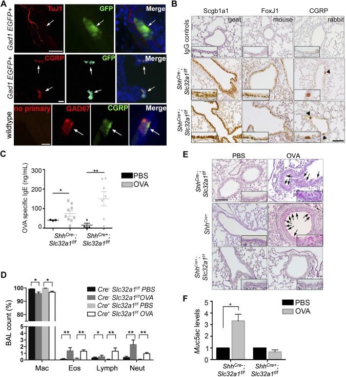
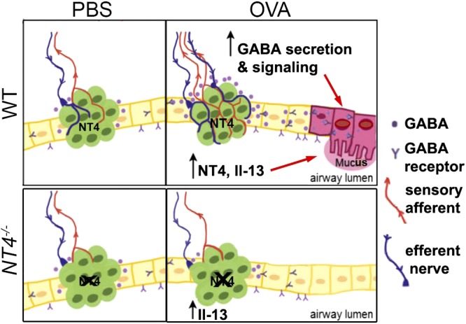
Pulmonary neuroendocrine cells (PNECs) are the only innervated airway epithelial cells. To what extent neural innervation regulates PNEC secretion and function is unknown. Here, we discover that neurotrophin 4 (NT4) plays an essential role in mucus overproduction after early life allergen exposure by orchestrating PNEC innervation and secretion of GABA. We found that PNECs were the only cellular source of GABA in airways. In addition, PNECs expressed NT4 as a target-derived mechanism underlying PNEC innervation during development. Early life allergen exposure elevated the level of NT4 and caused PNEC hyperinnervation and nodose neuron hyperactivity. Associated with aberrant PNEC innervation, the authors discovered that GABA hypersecretion was required for the induction of mucin Muc5ac expression. In contrast, NT4-/- mice were protected from allergen-induced mucus overproduction and changes along the nerve-PNEC axis without any defects in inflammation. Last, GABA installation restored mucus overproduction in NT4-/- mice after early life allergen exposure. Together, our findings provide the first evidence for NT4-dependent neural regulation of PNEC secretion of GABA in a neonatal disease model. Targeting the nerve-PNEC axis may be a valid treatment strategy for mucus overproduction in airway diseases, such as childhood asthma.-Barrios, J., Patel, K. R., Aven, L., Achey, R., Minns, M. S., Lee, Y., Trinkaus-Randall, V. E., Ai, X. Early life allergen-induced mucus overproduction requires augmented neural stimulation of pulmonary neuroendocrine cell secretion.

Keywords: GABA; NT4; childhood asthma; innervation.


Figures

Figure 1.
Figure 1.
NT4 was required for mucus overproduction after early life allergen exposure. A) Experimental scheme of early life OVA exposure in mice. Control mice were challenged with saline (PBS). Lungs were collected at P21. B) Serum levels of IL-13 measured by ELISA. Each mark represents 1 mouse. C) Differential immune cell counts in BAL. Relative abundance of macrophages (Mac), eosinophils (Eos), neutrophils (Neut), and lymphocytes (Lymph) was quantified as percentage of cells in BAL; n = 9 per group. D) Representative images of PAS staining in airways of WT and NT4−/− mice challenged with PBS or OVA. Mucin+ cells are marked by arrows. Enlarged epithelium staining is shown in inserts. E) Muc5ac mRNA expression in WT and NT4−/− mice that were challenged with PBS or OVA; n = 14 mice for each group. F) Muc5ac mRNA levels in lung slices of P21 WT mice (n = 3) after 48 h in culture. Results were normalized to PBS-challenged controls. G) Muc5ac mRNA levels in lung slices of P21 NT4−/− mice (n = 6) after 48 h in culture with and without neurotransmitter treatment. Results were normalized to PBS-challenged slices without treatment. Neurotransmitters tested included GABA (10 mg/ml), CGRP (10 µM), substance P (SP, 10 µM), dopamine (dopa, 10 µM), and Mch (10 μM). Data in C–E represent means ± sem from 3 independent experiments. *P < 0.05, **P < 0.01. Scale bar, 100 µm.
Figure 2.
Figure 2.
GABA from PNECs was required for mucus overproduction after early life OVA exposure. A) PNEC expression of GAD67 assayed at P21. In lungs of GAD1-GFP mice, PNECs were GFP+ while TuJ1+ nerves were not. Double staining for Gad67 and PNEC marker CGRP also showed colocalization. Arrows indicate PNECs. Scale bars, 25 µm. B) Characterization of airway epithelium in ShhCre−;Slc32a1f/f control littermates and ShhCre+;Slc32a1f/f mice at P21 by staining for specific markers of club cells (Scgb1a1), ciliated cells (Foxj1), and PNECs (CGRP). Negative controls were matched IgGs. Arrowheads indicate PNEC clusters. Insets show enlarged images of epithelium staining. Scale bars, 100 µm. C) Serum levels of OVA-specific IgE in saline and OVA-exposed littermates and ShhCre+;Slc32a1f/f mice at P21. Each mark indicates sample from 1 mouse. D) Differential immune cell count in BAL of PBS- and OVA-exposed control (Cre) and ShhCre+;Slc32a1f/f (Cre+) mice at P21. Percentages of macrophages (Mac), eosinophils (Eos), lymphocytes (Lymph), and neutrophils (Neut) are shown. E) Representative images of PAS staining in airways of ShhCre−;Slc32a1f/f and ShhCre+ controls and ShhCre+;Slc32a1f/f mutant mice at P21. Arrows indicate mucin-positive cells. Scale bars, 100 µm. Insets show enlarged images of stained epithelium. F) Muc5ac gene expression in lungs of littermate controls (PBS; n = 4; OVA, n = 11) and ShhCre+;Slc32a1f/f mutant mice (PBS, n = 6; OVA, n = 5). Data in C, D, and F represent means ± sem from 3 independent experiments. *P < 0.05, **P < 0.01.
Figure 3.
Figure 3.
PNEC innervation during postnatal development was dependent on NT4. A) Quantification of solitary PNECs, clusters, and total number of PNECs at birth and at P7, 14, and 21 in WT mice. PNECs were quantified after CGRP staining of tissue sections collected from entire right cranial lobe. B) PNEC expression of NT4 by double staining for NT4 and CGRP. NT4−/− lung sections were negative control for NT4 antibody. Arrows indicate PNECs. Scale bars, 100 µm. C) Representative images of postnatal TrkB lineage tracing using TrkBCreERT2/+;Rosa(tmRed) mouse line. PNECs (arrows) were stained for CGRP indicated by arrows. Nerves (arrowheads) were labeled by tomato red (tmRed) fluorescence. Scale bar, 100 µm. D) Assessment of solitary PNECs and clusters in WT and NT4−/− lungs with and without OVA challenges at P21. PNECs were labeled by CGRP staining of medial lung slices from left lobe. Each mark indicates results from 1 lung slice; n = 3 for each group. E) Assessment of NEB innervation in WT and NT4−/− mice with and without OVA exposure. Lung slices were stained with CGRP and TuJ1 antibodies followed by confocal microscopy to assess spatial association between NEBs and nerves. NEBs with 5 or more PNECs were counted. NEBs without associated nerves (none), nerves at base (basal), and nerves penetrating into clusters (penetrating) were grouped and presented as percentages of all NEBs. F) Representative images of NEBs with penetrating nerves in WT and NT4−/− mice with and without OVA challenges at P21. Scale bar, 50 µm. G) Quantification of NEB innervation density. Density of nerves was calculated by normalizing TuJ1-immunoreactive area by number of cells within NEBs in WT and NT4−/− mice with and without OVA challenges. For data in E–G, total of 45–50 NEBs in lung slices from 3 mice were scored for each group. N.s., not significant. *P < 0.05.
Figure 4.
Figure 4.
NT4 was required for elevated purinergic receptor gene expression and increased nodose ganglion neuronal activities after early life OVA exposure. A) Representative images of double staining of medial nodose ganglions in WT and NT4−/− mice at P21 for pan neuronal marker Uchl1 and P2X3 receptor. Nuclei were labeled by DAPI. Scale bars, 250 µm. B) Quantification of neuronal numbers in medial sections of nodose ganglions from WT and NT4−/− mice at P21. Eighteen sections from 6 ganglions of 3 mice were quantified. C) Quantification of percentages of P2X3+ neurons in nodose ganglions from WT and NT4−/− mice at P21. D) P2X3 mRNA levels in nodose ganglions from WT and NT4−/− mice with and without OVA exposure. Six ganglions from 3 mice were pooled as 1 sample. Three samples were analyzed for each condition. E) Representative images of P2X3 staining of primary nodose ganglion neurons after 72 h in culture treated with control BSA or NT4. Arrowheads indicate P2X3+ neurons. F) Ca2+ imaging of control BSA- and NT4-treated primary nodose ganglion neurons in response to ATP stimulation. Relative fluorescence (Ft/F0) of Fluo-3 AM before and after ATP stimulation (arrow) was measured. To block P2X3 activities, NT4-treated cultures were pretreated with A-317491 (1 µM) for 20 min before ATP stimulation and during entire course of Ca2+ imaging. Results represent 2 independent experiments. Data in B–D represent means ± sem of 3 independent experiments. **P < 0.01.
Figure 5.
Figure 5.
NT4 was required for GABA secretion and mucus overproduction after early life OVA exposure. A) Serum levels of GABA in WT and NT4−/− mice with and without OVA challenges measured by ELISA. Data represent means ± sem from 10 to 16 mice of each group. B) Gad1 mRNA expression in airway epithelium of WT and NT4−/− mice after PBS or OVA exposure at P21; n = 6. C, D) Serum levels of IgE (C) and IL-13 (D) in BAL of PBS-exposed NT4−/− mice and OVA-exposed NT4−/− mice with and without intratracheal GABA treatment. Each mark indicates sample from 1 mouse. E) Representative images of PAS staining of lungs sections of WT and NT4−/− mice with indicated treatment. Arrows indicate mucin+ cells. Insets show enlarged images of epithelium staining. F) Muc5ac gene expression in PBS- and OVA-exposed WT and NT4−/− mice with and without GABA treatment. Each group had minimum of 7 mice. Data represent means ± sem of at least 3 independent experiments. G) Foxa3 staining of lung sections of WT and NT4−/− mice with and without OVA exposure and GABA treatment. Arrows indicate Foxa3+ cells. H) Percentages of Foxa3+ cells in airway epithelium of each experimental group. Data represent means ± sem from 15 airways of 3 mice for each experimental condition. Scale bars, 100 μm. *P < 0.05, **P < 0.01, ***P < 0.001.
Figure 6.
Figure 6.
Model of NT4-PNEC axis in GABA secretion and mucus overproduction after early life allergen exposure. PNECs are innervated by both sensory afferents and efferent nerves. During development, NT4 expressed by PNECs serves as target-derived trophic factor for innervating nerves to establish innervation. Allergen exposure during postnatal PNEC development elevates levels of NT4, which in turn causes PNEC hyperinnervation and hyperactive nodose sensory neurons that feed on efferent nerves. Hyperactive neurocircuitry deregulates GABA secretion from PNECs. Aberrant levels of GABA act together with IL-13 to induce mucus overproduction. Loss of NT4 reduces PNEC innervation at baseline and prevents allergen-induced PNEC hyperinnervation. There is thus no induction of GABA secretion from PNECs in allergen-exposed NT4−/− mice. IL-13, in absence of GABA secretion, fails to induce mucus overproduction.

References

    1. Ordoñez C. L., Khashayar R., Wong H. H., Ferrando R., Wu R., Hyde D. M., Hotchkiss J. A., Zhang Y., Novikov A., Dolganov G., Fahy J. V. (2001) Mild and moderate asthma is associated with airway goblet cell hyperplasia and abnormalities in mucin gene expression. Am. J. Respir. Crit. Care Med. 163, 517–523 - PubMed
    1. Evans C. M., Williams O. W., Tuvim M. J., Nigam R., Mixides G. P., Blackburn M. R., DeMayo F. J., Burns A. R., Smith C., Reynolds S. D., Stripp B. R., Dickey B. F. (2004) Mucin is produced by clara cells in the proximal airways of antigen-challenged mice. Am. J. Respir. Cell Mol. Biol. 31, 382–394 - PMC - PubMed
    1. Pardo-Saganta A., Law B. M., Gonzalez-Celeiro M., Vinarsky V., Rajagopal J. (2013) Ciliated cells of pseudostratified airway epithelium do not become mucous cells after ovalbumin challenge. Am. J. Respir. Cell Mol. Biol. 48, 364–373 - PMC - PubMed
    1. Wills-Karp M., Luyimbazi J., Xu X., Schofield B., Neben T. Y., Karp C. L., Donaldson D. D. (1998) Interleukin-13: central mediator of allergic asthma. Science 282, 2258–2261 - PubMed
    1. Chen G., Korfhagen T. R., Xu Y., Kitzmiller J., Wert S. E., Maeda Y., Gregorieff A., Clevers H., Whitsett J. A. (2009) SPDEF is required for mouse pulmonary goblet cell differentiation and regulates a network of genes associated with mucus production. J. Clin. Invest. 119, 2914–2924 - PMC - PubMed

Publication types