. 2017 May 31;7(1):2525.
doi: 10.1038/s41598-017-02890-y.

Colitis promotes neuronal differentiation of Sox2+ and PLP1+ enteric cells

Affiliations

Colitis promotes neuronal differentiation of Sox2+ and PLP1+ enteric cells

Jaime Belkind-Gerson et al. Sci Rep. .

Abstract

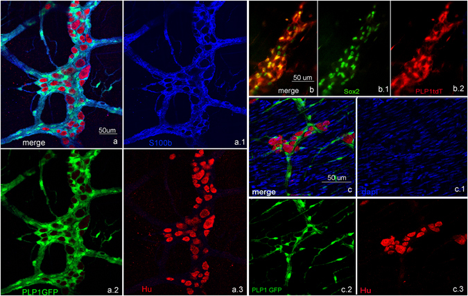
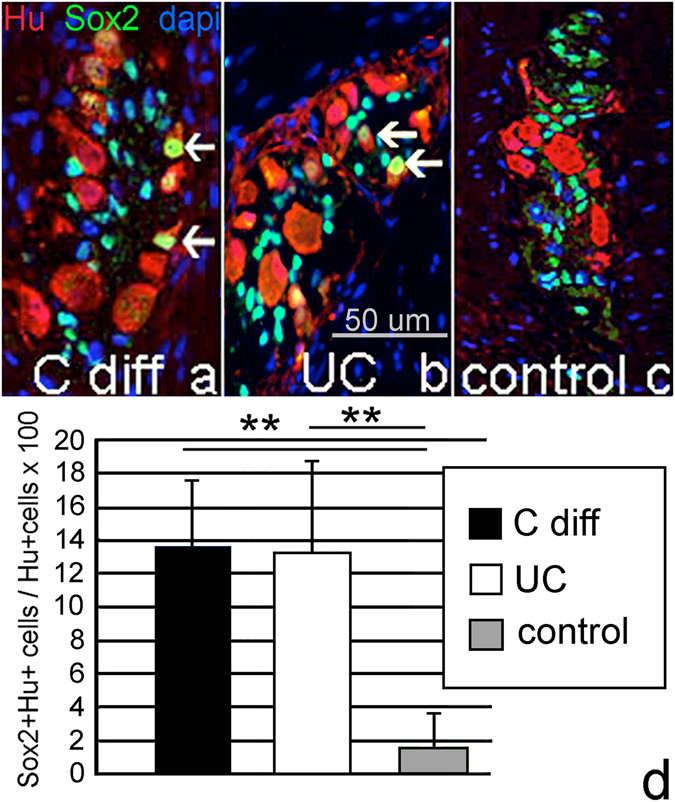
Mechanisms mediating adult enteric neurogenesis are largely unknown. Using inflammation-associated neurogenesis models and a transgenic approach, we aimed to understand the cell-source for new neurons in infectious and inflammatory colitis. Dextran sodium sulfate (DSS) and Citrobacter rodentium colitis (CC) was induced in adult mice and colonic neurons were quantified. Sox2GFP and PLP1GFP mice confirmed the cell-type specificity of these markers. Sox2CreER:YFP and PLP1creER:tdT mice were used to determine the fate of these cells after colitis. Sox2 expression was investigated in colonic neurons of human patients with Clostridium difficile or ulcerative colitis. Both DSS and CC led to increased colonic neurons. Following colitis in adult Sox2CreER:YFP mice, YFP initially expressed predominantly by glia becomes expressed by neurons following colitis, without observable DNA replication. Similarly in PLP1CreER:tdT mice, PLP1 cells that co-express S100b but not RET also give rise to neurons following colitis. In human colitis, Sox2-expressing neurons increase from 1-2% to an average 14% in colitis. The new neurons predominantly express calretinin, thus appear to be excitatory. These results suggest that colitis promotes rapid enteric neurogenesis in adult mice and humans through differentiation of Sox2- and PLP1-expressing cells, which represent enteric glia and/or neural progenitors. Further defining neurogenesis will improve understanding and treatment of injury-associated intestinal motility/sensory disorders.


Conflict of interest statement

The authors declare that they have no competing interests.

Figures

Figure 1
Figure 1
Chemical and infectious colitis both promote enteric neurogenesis. DSS colitis (n = 8) and Citrobacter rodentium colitis (n = 8) were induced in 4 month-old mice and the colons processed for immunohistochemistry using Hu antibody to label enteric neurons. Significantly more neurons are present throughout the colon in both DSS (a,b) and Citrobacter rodentium (f,g) models. This increased neuronal density is associated with an increase in the proportion of Hu+ neurons that label with Sox2 (c-e and h-j, arrows point to Hu + Sox2+ neurons in (d,e and j). *p < 0.05, **p < 0.01. (d and e) were taken at 200x, (i and j) at 400x).
Figure 2
Figure 2
Sox2, PLP1 and S100b label enteric glia. Sox2GFP (a) labels most enteric glial cells in the adult mouse colon. Similarly, PLP1GFP (b) also labels most enteric glial cells. S100b is a third marker labeling most glial cells (c). GFAP, however only labels a fraction of enteric glia (d,d.3 close up of boxed area in d). (All Figures were taken at 200x, except closeup maginfications).
Figure 3
Figure 3
Colitis induces differentiation of Sox2+ glial cells to enteric neurons. Sox2 is normally expressed in the adult colon in glia both in the SOX2GFP mouse (a) as well as using anti-Sox2 antibodies (Sox2(ab)), as shown in (b). After 4 months of age, only occasional neurons express Sox2 protein (b, arrow). At 2 months of age, 3.5% of Hu+ colonic neurons express YFP (c, thick arrow), but in the Sox2CreER as in the other mouse models, these are rare by 4 months, when Sox2 expression is primarily limited to glia (d). After tamoxifen, DSS was given to 4 month-old mice for 7 days, followed by sacrifice on day 10. Treatment with DSS leads to a significant increase in YFP-expressing Hu+ neurons (e), thick arrows; thin arrows point to YFP+ glial cells). YFP+ neurons are also positive for PGP9.5 (f). In 4 month-old mice, the proportion of Hu+ neurons that express YFP increases from 1.1 ± 0.6% in controls to 4.4% ± 1.9% after colitis (g). A similar increase is observed when tamoxifen is administered 3 weeks before DSS, ensuring that residual tamoxifen is no longer present, 0.7% ± 0.2 in control and 2.4% ± 0.1 after DSS (h), (**p < 0.01) n = 4. (a,c,e and f) were taken at 200x, (b and d) at 100x).
Figure 4
Figure 4
Glia give rise to neurons without new DNA synthesis. EdU was administered i.p. daily (50 mg/Kg) to Sox2creER:YFP mice receiving DSS for 7 days and examined 2 days later. EdU incorporation was seen in the mucosa (muc a, a.1), but not in Hu+ or Hu + YFP+ neurons (arrow). (b) Magnified view of myenteric ganglia. (Fig. 4 was taken at 200X, except closeup magnification 4.b).
Figure 5
Figure 5
PLP1 is expressed by enteric glia and not neurons. PLP1tdT+ cells co-express S100b (a). PLP1tdT expressing cells also express Sox2 (b). In PLP1GFP mice, enteric neurons are not GFP+ in the adult gut (c). (All images of Fig. 5 were taken at 200X).
Figure 6
Figure 6
PLP1+ enteric glia give rise to neurons in vitro and ex vivo. Enteric neurospheres derived from PLP1CreER:tdT mice contain >90% tdT+ cells by confocal imaging (a,b close up of boxed area in a). The majority of cells dissociated from these neurospheres are tdT+ (c). Following culture, PLP1CreER:tdT-derived cells are positive for Hu and Sox2 (d), PGP9.5 and Sox2 (e), Tuj1 and Sox2, ((f) blue arrows denote triple-expressing cells), and S100b and Sox2 (g). Cultured colonic LMMP from PLP1CreER:tdT mice also shows tdT+/Hu+ double-expressing cells (h, arrows). All images of Fig. 6 were taken at 100X except closeup. (5b and f,h at 200x).
Figure 7
Figure 7
PLP1+ enteric glia give rise to neurons after colitis. PLP1CreER:tdT is not normally expressed in neurons in adult mice (a). However, mice give rise to Hu+ neurons following colitis in the submucosal (a.1) and myenteric plexus (a.2,b–b.3, arrows point to PLP1tdT + Hu+ cells). In normal, non-inflamed colon, glial cells do not express RET, which is normally expressed by neurons and progenitors (cc.3). Similarly, Sox2+ cells do not co-express RET in the adult ENS (d, arrows), nor do PLP1+ cells (ee2). (Images 7.a, and 7.c were taken at 200x except for closeups, 7.be were taken at 400X).
Figure 8
Figure 8
Colitis induces YFP + Hu+ cells that subsequently downregulate Sox2 expression. In the myenteric ganglia of control mice, all YFP + Hu+ cells (YFP green, Hu white) express Sox2 (red) (aa.4 arrow). Following DSS 50% of the YFP+ neurons no longer express Sox2 protein (bb.4 arrowheads). A 25% increase in Hu + YFP + Sox2+ cells is still observed in DSS-treated mice, (c, blue bar), but many of the new Hu+ neurons are negative for Sox2 protein expression (c, green bar). *p < 0.05. (All Images were taken at 200x).
Figure 9
Figure 9
Infectious and inflammatory colitis in humans leads to an expansion of Sox2 + Hu+ neurons. Patients with Clostridium difficile (n = 15) (a) or ulcerative colitis (n = 15) (b) have significantly more Sox2 + Hu+ neurons than control colon (c), where Sox2 + Hu+ neurons are infrequently observed (d). (All Images were taken at 200x).
Figure 10
Figure 10
Newly formed neurons following colitis are predominantly calretinin excitatory motor neurons, which form synapses. In the myenteric ganglia of control mice, approximately 25% of Hu+ neurons express calretinin ((a) Hu green, calretinin red, arrows point to double-expressing cells). In our model, however 82% of YFP+ neurons (thus the newly formed neurons) express calretinin (bb.4 YFP green, calretinin white, Hu red. Arrows in b point to triple-expressing cells). nNOS is normally expressed by 19% of Hu+ neurons in control ((c) nNOS green, Hu red. Arrows point to double-expressing cells), but was only found to be expressed by 13% of YFP+ neurons post-colitis (d.1d.4 YFP green, Hu red and nNOS white. Arrows point to triple-expressing cells). The newly formed neurons synthesize synapses as manifested by synaptophysin expression (e). *p < 0.05, **p < 0.01. (All Images were taken at 200x).
Figure 11
Figure 11
Inflammation induced early neurogenesis. In the present work, we show that inflammation can cause a rapid glial-to-neuronal fate change or neural progenitor differentiation, which can be identified by an expanding neuronal mass and a new population of Sox2 + Hu+ neurons. We hypothesize that these then downregulate Sox2 as they become mature neurons.

References

    1. Srinivasan, S. & Wiley, J. W. New insights into neural injury, repair, and adaptation in visceral afferents and the enteric nervous system. Curr Opin Gastroenterol16, 78–82, doi:00001574-200001000-00014 [pii] (2000). - PubMed
    1. Becker, L., Peterson, J., Kulkarni, S. & Pasricha, P. J. Ex vivo neurogenesis within enteric ganglia occurs in a PTEN dependent manner. PLoS One8, e59452, doi:10.1371/journal.pone.0059452 PONE-D-12-30691 [pii] (2013). - PMC - PubMed
    1. Urban N, Guillemot F. Neurogenesis in the embryonic and adult brain: same regulators, different roles. Front Cell Neurosci. 2014;8:396. doi: 10.3389/fncel.2014.00396. - DOI - PMC - PubMed
    1. Liu MT, Kuan YH, Wang J, Hen R, Gershon MD. 5-HT4 receptor-mediated neuroprotection and neurogenesis in the enteric nervous system of adult mice. J Neurosci. 2009;29:9683–9699. doi: 10.1523/JNEUROSCI.1145-09.2009. - DOI - PMC - PubMed
    1. Laranjeira C, et al. Glial cells in the mouse enteric nervous system can undergo neurogenesis in response to injury. J Clin Invest. 2011;121:3412–3424. doi: 10.1172/JCI58200. - DOI - PMC - PubMed

MeSH terms