. 2017 May-Jun;51(3):256-268.
doi: 10.4103/ortho.IJOrtho_199_16.

Infected nonunion of tibia

Affiliations

Infected nonunion of tibia

Milind Madhav Chaudhary. Indian J Orthop. 2017 May-Jun.

Abstract

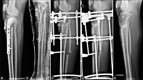
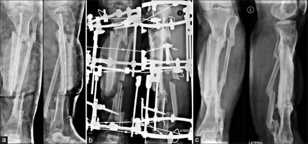
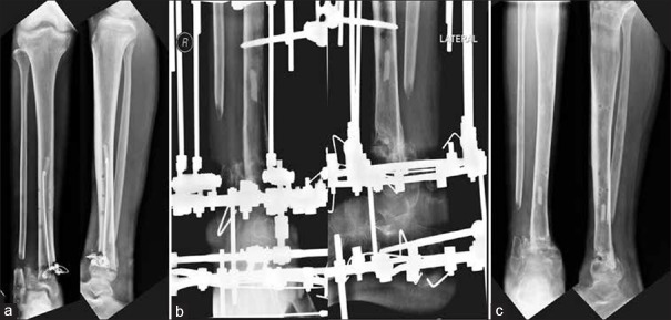
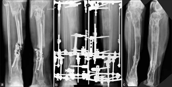
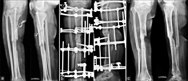
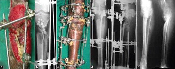
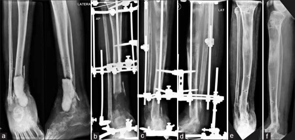
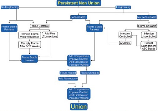
Infected nonunions of tibia pose many challenges to the treating surgeon and the patient. Challenges include recalcitrant infection, complex deformities, sclerotic bone ends, large bone gaps, shortening, and joint stiffness. They are easy to diagnose and difficult to treat. The ASAMI classification helps decide treatment. The nonunion severity score proposed by Calori measures many parameters to give a prognosis. The infection severity score uses simple clinical signs to grade severity of infection. This determines number of surgeries and allows choice of hardware, either external or internal for definitive treatment. Co-morbid factors such as smoking, diabetes, nonsteroidal anti-inflammatory drug use, and hypovitaminosis D influence the choice and duration of treatment. Thorough debridement is the mainstay of treatment. Removal of all necrotic bone and soft tissue is needed. Care is exercised in shaping bone ends. Internal fixation can help achieve union if infection was mild. Severe infections need external fixation use in a second stage. Compression at nonunion site achieves union. It can be combined with a corticotomy lengthening at a distant site for equalization. Soft tissue deficit has to be covered by flaps, either local or microvascular. Bone gaps are best filled with the reliable technique of bone transport. Regenerate bone may be formed proximally, distally, or at both sites. Acute compression can fill bone gaps and may need a fibular resection. Gradual reduction of bone gap happens with bone transport, without need for fibulectomy. When bone ends dock, union may be achieved by vertical or horizontal compression. Biological stimulus from iliac crest bone grafts, bone marrow aspirate injections, and platelet concentrates hasten union. Bone graft substitutes add volume to graft and help fill defects. Addition of rh-BMP-7 may help in healing albeit at a much higher cost. Regeneration may need stimulation and augmentation. Induced membrane technique is an alternative to bone transport to fill gaps. It needs large amounts of bone graft from iliac crest or femoral canal. This is an expensive method physiologically and economically. Infection can resorb the graft and cause failure of treatment. It can be done in select cases after thorough eradication of infection. Patience and perseverance are needed for successful resolution of infection and achieving union.

Keywords: Bone cements; Infection; antibiotic cement; bone diseases; bone regeneration; bone transport; gap nonunion; induced membrane technique; infection severity score; infections; nonunion; regenerate bone; tibia.


Conflict of interest statement

There are no conflicts of interest.

Figures

Figure 1
Figure 1
Oblique view of leg bones with knee showing (a) upper tibial infected nonunion (b) After infection control (c) Ilizarov apparatus in situ (d). If vertical compression is given, it causes vertical displacement of nonunion ends. With the help of washers, horizontal compression achieved perpendicular to plane of nonunion (e) Good union. No loss of length
Figure 2
Figure 2
X-ray leg bones with knee and ankle joint showing (a) Infected segmental nonunion after cure of infection with rods and beads (b) Ilizarov fixator in situ to achieve horizontal compression perpendicular to plane of nonunion (c) achieved union at both levels. Fibulaectomy done. No lengthening
Figure 3
Figure 3
X-ray anteroposterior and lateral views with knee and ankle joints showing (a) Infected nonunion lower tibia. Beads inserted (b) Ilizarov fixator applied. Acute compression at nonunion site done gradually at low rate hence no angular deformity at nonunion site despite an irregular shaped defect. Simultaneous lengthening at upper level to equalize lengths (c) Union achieved at distal end with good regenerate at proximal corticotomy site and no limb length discrepancy
Figure 4
Figure 4
X-ray anteroposterior and lateral views showing (a) Infected gap in M3 tibia. Very poor fibrotic soft tissues. Free flap failed because recipient vessel was not found intact (b) Proximal tibial bone transport. Docking and compression without bone grafting (c) Using ilizarov technique achieved union. Good alignment
Figure 5
Figure 5
X-ray leg bones with knee joint anteroposterior and lateral views showing (a) Infected tibial nonunion. Irregular bony ends. Had we resected bone till achieving horizontal bony surfaces, it would have created a lot of shortening (b) we gave compression at rate of one-fourth mm twice a week to achieve compression but not cause an angular deviation. No bone grafting done (c) Sound union, after ilizarov construct removal
Figure 6
Figure 6
(a) Clinical photograph showing severe soft tissue loss and bone loss in upper tibia. Debridement and beads seen. Inferiorly based fasciocutaneous flap was done for cover. X-ray leg bones with knee joint anteroposterior view (c) Clinical photograph showing full stent of frame across the knee. Femur included in frame for stability. (d) X-ray anteroposterior and lateral views showing that distal tibial corticotomy done for filling gap (e) X-ray anteroposterior and lateral views of leg bones with knee and ankle joint showing sound union. No deformity, no limb length discrepancy (LLD) and no infection persists
Figure 7
Figure 7
(a) X-ray leg bones with ankle joint anteriposterior and lateral views showing large gap after debridement. Cement block used (b-d) X-ray anterioposterior and lateral views showing reamer irrigator-aspirator graft inserted after nucleating cement block from membrane at 4 weeks and membrane sutured. Graft completely resorbed due to mild recrudescence of infection. Problem solved with Proximal to distal bone transport. Bone transport is most reliable method. Masquelet method may not work in all cases (e and f) X-ray anteroposterior and lateral views showing sound union
Chart 1
Chart 1
Algorithm for dealing with persistent nonunion while in the Ilizarov fixator. This can be a common problem. Augmentation of fixation is needed frequently with new pins. Fistulectomy helps achieve proper compression at Docking site or nonunion site. Biological augmentation is bone grafts, bone marrow injections, platelet concentrates and BG substitutes. BMP, if affordable

References

    1. Keating JF, Simpson AH, Robinson CM. The management of fractures with bone loss. J Bone Joint Surg Br. 2005 Feb;87(2):142–50. - PubMed
    1. Bose D, Kugan R, Stubbs D, McNally M. Management of infected nonunion of the long bones by a multidisciplinary team. Bone Joint J. 2015;97-B:814–7. - PubMed
    1. Dendrinos GK, Kontos S, Lyritsis E. Use of the Ilizarov technique for treatment of nonunion of the tibia associated with infection. J Bone Joint Surg Am. 1995;77:835–46. - PubMed
    1. Mora R, Pedrotti L, Bertani B, Quattrini F. Treatment of infected nonunions. In: Mora R, editor. Nonunion of the Long Bones: Diagnosis and Treatment with Compression-Distraction Techniques. New York: Springer; 2006. pp. 199–223.
    1. Borzunov DY, Balaev PI, Subramanyam KN. Reconstruction by bone transport after resection of benign tumors of tibia: A retrospective study of 38 patients. Indian J Orthop. 2015;49:516–22. - PMC - PubMed

LinkOut - more resources