Aberrant right subclavian artery presenting as tracheoesophagial fistula in a 50-year-old lady: Case report of a rare presentation of a common arch anomaly
- PMID: 28566828
- PMCID: PMC5431032
- DOI: 10.4103/apc.APC_158_16
Aberrant right subclavian artery presenting as tracheoesophagial fistula in a 50-year-old lady: Case report of a rare presentation of a common arch anomaly
Abstract
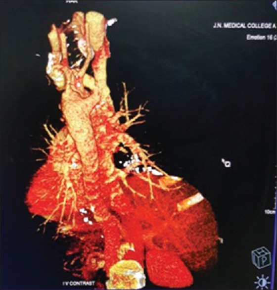
A 50-year-old, woman with a 2-year history of progressive dysphagia and 2-month history of chronic cough was referred to our center in a state of generalized sepsis. Provisional diagnosis of carcinoma esophagus with tracheoesophagial fistula was made. Evaluation of the patient revealed an aberrant right subclavian artery with retroesophageal course with compression of the esophagus and trachea with fistulous communication in between. The patient was managed with medical stabilization and with feeding jejunostomy, but she succumbed to underlying severe sepsis. This presentation of aberrant subclavian artery at this advanced age rare and is therefore reported.
Keywords: Aberrant right subclavian artery; aspiration pneumonitis; tracheoesophageal fistula.
Conflict of interest statement
There are no conflicts of interest.
Figures


References
-
- Richardson JV, Doty DB, Rossi NP, Ehrenhaft JL. Operation for aortic arch anomalies. Ann Thorac Surg. 1981;31:426–32. - PubMed
-
- De Luca L, Bergman JJ, Tytgat GN, Fockens P. EUS imaging of the arteria lusoria: Case series and review. Gastrointest Endosc. 2000;52:670–3. - PubMed
-
- Stone WM, Brewster DC, Moncure AC, Franklin DP, Cambria RP, Abbott WM. Aberrant right subclavian artery: Varied presentations and management options. J Vasc Surg. 1990;11:812–7. - PubMed
-
- Brown DL, Chapman WC, Edwards WH, Coltharp WH, Stoney WS. Dysphagia lusoria: Aberrant right subclavian artery with a Kommerell's diverticulum. Am Surg. 1993;59:582–6. - PubMed
Publication types
LinkOut - more resources
Full Text Sources
Other Literature Sources