

doi: 10.5021/ad.2017.29.3.365.
Epub 2017 May 11.
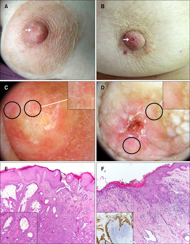
Dermoscopy as a Supportive Instrument in the Early Recognition of Erosive Adenomatosis of the Nipple and Mammary Paget's Disease
Affiliations
- PMID: 28566922
- PMCID: PMC5438952
- DOI: 10.5021/ad.2017.29.3.365
Item in Clipboard
Dermoscopy as a Supportive Instrument in the Early Recognition of Erosive Adenomatosis of the Nipple and Mammary Paget's Disease
Ann Dermatol.
2017 Jun.
No abstract available
Conflict of interest statement
CONFLICTS OF INTEREST: The authors have nothing to disclose.
Figures
References
-
- Takashima S, Fujita Y, Miyauchi T, Nomura T, Nishie W, Hamaoka H, et al. Dermoscopic observation in adenoma of the nipple. J Dermatol. 2015;42:341–342. - PubMed
-
- Errichetti E, Stinco G. The practical usefulness of dermoscopy in general dermatology. G Ital Dermatol Venereol. 2015;150:533–546. - PubMed
-
- Yanagishita T, Tamada Y, Tanaka M, Kasugai C, Takahashi E, Matsumoto Y, et al. Pigmented mammary Paget disease mimicking melanoma on dermatoscopy. J Am Acad Dermatol. 2011;64:e114–e116. - PubMed
-
- Brugués A, Iranzo P, Díaz A, Peña A, Estrach MT, Carrera C. Pigmented mammary Paget disease mimicking cutaneous malignant melanoma. J Am Acad Dermatol. 2015;72:e97–e98. - PubMed
LinkOut - more resources
Full Text Sources
Other Literature Sources
