. 2017 May 17:11:239.
doi: 10.3389/fnins.2017.00239. eCollection 2017.

Relative Contribution of Prolyl Hydroxylase-Dependent and -Independent Degradation of HIF-1alpha by Proteasomal Pathways in Cerebral Ischemia

Affiliations

Relative Contribution of Prolyl Hydroxylase-Dependent and -Independent Degradation of HIF-1alpha by Proteasomal Pathways in Cerebral Ischemia

Yomna Badawi et al. Front Neurosci. .

Abstract

Hypoxia inducible factor-1 (HIF-1) is a key regulator in hypoxia and can determine the fate of brain cells during ischemia. However, the mechanism of HIF-1 regulation is still not fully understood in ischemic brains. We tested a hypothesis that both the 26S and the 20S proteasomal pathways were involved in HIF-1α degradation under ischemic conditions. Using in vitro ischemic model (oxygen and glucose deprivation) and a mouse model of middle cerebral artery occlusion, we tested effects of inhibitors of proteasomes and prolyl hydroxylase (PHD) on HIF-1α stability and brain injury in cerebral ischemia. We observed that 30 and 60 min of oxygen-glucose deprivation significantly increased the 20S proteasomal activity. We demonstrated that proteasome inhibitors increased HIF-1α stabilization and cell viability and were more effective than PHD inhibitors in primary cultured cortical neurons exposed to oxygen and glucose deprivation. Furthermore, the administration of the proteasome inhibitor, epoxomicin, to mice resulted in smaller infarct size and brain edema than a PHD inhibitor. Our results indicate that 20S proteasomes are involved in HIF-1α degradation in ischemic neurons and that proteasomal inhibition provides more HIF-1α stabilization and neuroprotection than PHD inhibition in cerebral ischemia.

Keywords: HIF-1; neurons; oxidative stress; proteasome; stroke.


Figures

Figure 1
Figure 1
HIF-1α degradation by the 20S proteasomes. Immunoblotting for HIF-1α after the HIF-1α protein-beads were incubated with a cytosol fraction from SH-SY5Y cells and either the 26S or 20S proteasome for 1 and 3 h with the addition of (A) H2O2 or (B) ubiquitin.
Figure 2
Figure 2
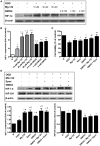
HIF-1α protein stabilization with the treatments of proteasome or PHD inhibitors. (A) Immunoblotting showing HIF-1α protein levels in neurons and (B) the quantitative results for Western blot data. Equalization of protein loading was determined using β-actin as the housekeeping protein. *p < 0.05 vs. normoxia (N), p < 0.05 vs. oxygen/glucose deprivation (OGD) (n = 3). (C) Neuronal viability assessed using the MTT assay *p < 0.05 vs. OGD, #p < 0.05 vs. DMOG (n = 3). (D) Immunoblotting showing HIF-1α and hydroxyl-HIF-1α (HIF-OH) protein levels in neurons. Equalization of protein loading was determined using β-actin as the housekeeping protein. Quantitative results for Western blot data. *p < 0.05 vs. N, #p < 0.05 vs. OGD (n = 3).
Figure 3
Figure 3
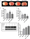
Effect of DMOG and epoxomicin (Epox) on HIF-1α expression and brain damage induced by cerebral ischemia. Brain damage as determined by TCC staining after mice were subjected to 90 min ischemia followed by 24 h reperfusion. (A) Representative TTC staining of brain coronal sections taken from the 3 mm position of the frontal pole. (B) Quantification of infarct volume determined by TTC stained sections (n = 5). Data presented as means ± SEM *p < 0.05, vs. control untreated mice. #p < 0.05, vs. mice treated with Epox. (C) Quantification of brain edema volume estimated from TTC stained sections (n = 5). Data presented as means ± SEM *p < 0.05, vs. control untreated mice. (D) Immunoblotting showing the levels of HIF-1α from the ipsilateral brain hemispheres of untreated mice and mice treated with dimethyloxalylglycine (DMOG), Epox or a combination of both (n = 3).
Figure 4
Figure 4
Schematic diagram of HIF-1α degradation mechanisms. Under normoxic conditions, HIF-1α is hydroxylated by prolyl hydroxylases (PHD), which targets it for degradation via the 26S proteasome pathway. During conditions of low oxygen and high oxidative stress, HIF-1α can be oxidized and subsequently degraded by the 20S proteasome.

References

    1. Baranova O., Miranda L. F., Pichiule P., Dragatsis I., Johnson R. S., Chavez J. C. (2007). Neuron-specific inactivation of the hypoxia inducible factor 1 increases brain injury in a mouse model of transient focal cerebral ischemia. J. Neurosci. 27:6320. 10.1523/JNEUROSCI.0449-07.2007 - DOI - PMC - PubMed
    1. Berti R., Williams A. J., Velarde L. C., Moffett J. R., Elliott P. J., Adams J., et al. . (2003). Effect of the proteasome inhibitor MLN519 on the expression of inflammatory molecules following middle cerebral artery occlusion and reperfusion in the rat. Neurotox. Res. 5, 505–514. 10.1007/BF03033160 - DOI - PubMed
    1. Breusing N., Grune T. (2008). Regulation of proteasome-mediated protein degradation during oxidative stress and aging. Biol. Chem. 389, 203–209. 10.1515/BC.2008.029 - DOI - PubMed
    1. Clark W. M., Lessov N. S., Dixon M. P., Eckenstein F. (1997). Monofilament intraluminal middle cerebral artery occlusion in the mouse. Neurol. Res. 19:641. 10.1080/01616412.1997.11740874 - DOI - PubMed
    1. Coux O., Tanaka K., Goldberg A. L. (1996). Structure and functions of the 20S and 26S proteasomes. Annu. Rev. Biochem. 65, 801–847. 10.1146/annurev.bi.65.070196.004101 - DOI - PubMed

LinkOut - more resources