

Imaging Brain Function with Functional Near-Infrared Spectroscopy in Unconstrained Environments
- PMID: 28567011
- PMCID: PMC5434677
- DOI: 10.3389/fnhum.2017.00258
Imaging Brain Function with Functional Near-Infrared Spectroscopy in Unconstrained Environments
Abstract
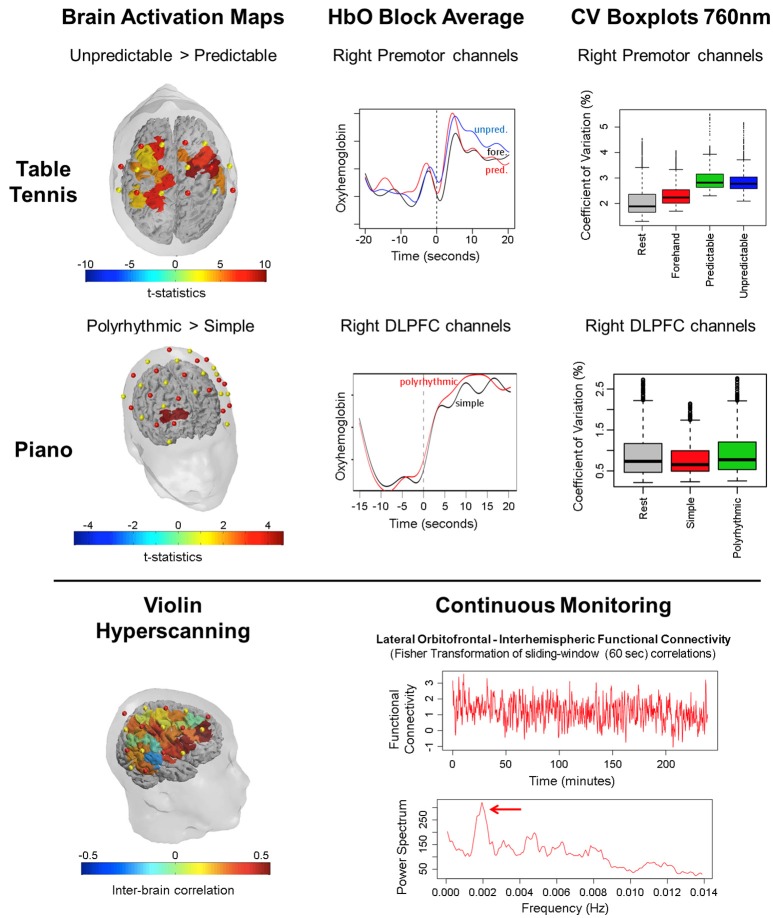
Assessing the neural correlates of motor and cognitive processes under naturalistic experimentation is challenging due to the movement constraints of traditional brain imaging technologies. The recent advent of portable technologies that are less sensitive to motion artifacts such as Functional Near Infrared Spectroscopy (fNIRS) have been made possible the study of brain function in freely-moving participants. In this paper, we describe a series of proof-of-concept experiments examining the potential of fNIRS in assessing the neural correlates of cognitive and motor processes in unconstrained environments. We show illustrative applications for practicing a sport (i.e., table tennis), playing a musical instrument (i.e., piano and violin) alone or in duo and performing daily activities for many hours (i.e., continuous monitoring). Our results expand upon previous research on the feasibility and robustness of fNIRS to monitor brain hemodynamic changes in different real life settings. We believe that these preliminary results showing the flexibility and robustness of fNIRS measurements may contribute by inspiring future work in the field of applied neuroscience.
Keywords: brain imaging; continuous monitoring; fNIRS; hyperscanning; musicians; naturalistic experimentation; sports; wearable.
Figures

References
-
- Ayaz H., Onaral B., Izzetoglu K., Shewokis P. A., McKendrick R., Parasuraman R. (2013). Continuous monitoring of brain dynamics with functional near infrared spectroscopy as a tool for neuroergonomic research: empirical examples and a technological development. Front. Hum. Neurosci. 7:871. 10.3389/fnhum.2013.00871 - DOI - PMC - PubMed
LinkOut - more resources
Full Text Sources
Other Literature Sources