

Review
doi: 10.2174/1874357901711010015.
eCollection 2017.
Cytomegalovirus, Macrophages and Breast Cancer
Affiliations
- PMID: 28567162
- PMCID: PMC5420183
- DOI: 10.2174/1874357901711010015
Item in Clipboard
Review
Cytomegalovirus, Macrophages and Breast Cancer
Open Virol J.
.
Abstract
The human cytomegalovirus (HCMV) is a betaherpesvirus that is highly host specific, infects among others epithelial cells and macrophages, and has been recently mentioned as having oncomodulatory properties. HCMV is detected in the breast tumor tissue where macrophages, especially tumor associated macrophages, are associated with a poor prognosis. In this review, we will discuss the potential implication of HCMV in breast cancer with emphasis on the role played by macrophages.
Keywords: Breast cancer; HCMV; Macrophages; Tumor.
Figures
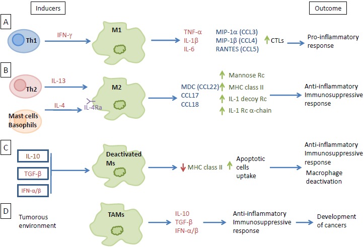
Macrophages, heterogeneous cell populations. (A) Th1 induction of M1 through interferon-gamma signaling. M1 releases specific cytokines and chemokines as part of the proinflammatory response. (B) M2 differentiation via IL-13 and IL-4 released from Th2, mast cells, and basophils. Differentiated M2 then releases factors that favor the anti-inflammatory and immunosuppressive responses. (C) Ms deactivation through the IL-10, TGF-beta, and IFN-alpha/beta signaling. Deactivation downregulates the expression of MHC class II and increases the uptake of apoptotic cells, resulting in an anti-inflammatory and immunosuppressive response. (D) Macrophages differentiation towards a specific phenotype present in the tumorous environment (TAMs), where they release large amount of immunosuppressive cytokines and a little amount of pro-inflammatory cytokines indirectly promoting the development of cancer.
CTLs: Th1-cytotoxic T cells ; IFN: Interferon ; IL: Interleukine ; MHC: Major Histocompatibility Complex ; MIP: Macrophage Inflammatory Protein ; TNF: Tumor Necrosis Factor ; TAMs: Tumor Associated Macrophages ; Th: T helper ; Rc: Receptor ; CCL : C-C Motif Chemokine Ligand ; M : Macrophage ; MDC : Macrophage-Derived Chemokine ; RANTES : Regulated on Activation, Normal T cell Expressed and Secreted ; TGF : Transforming Growth Factor.
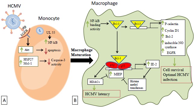
Cellular transduction events occuring after macrophages infection by HCMV. (A) HCMV binds to the cellular EGFR through its glycoprotein gB and directly activates Akt, thus promoting an Akt-dependent prosurvival state. EGFR activation rapidly induces the expression of HSP27 and Mcl-1 that act together to downregulate caspase-3 activity and permit the virus persistence. (B) In the differentiated macrophages, NF-kB binding activity is increased allowing Bcl-3 to activate a number of human genes that favor cellular survival and optimal infection by HCMV. Activated p52/Bcl-3 complexes also act through the NF-kB sites to regulate the MIEP of HCMV and thus affecting the virus latency via HDACs. MIEP is also under by negative autoregulation through IE2 expression which in turn activates histone methyltransferases.
EGFR: Epidermal Growth Factor Receptor ; HDACs: Histone Deacetylases ; HCMV: Human Cytomegalovirus ; MIEP: Major Immediate Early Promoter ; Bcl: B-cell lymphoma encoded protein ; gB: Glycoprotein B ; HSP: Heat Shock Protein ; IE: Immediate Early ; Mcl-1: Induced myeloid leukemia cell differentiation protein ; NF-kB: Nuclear Factor-Kappa B.
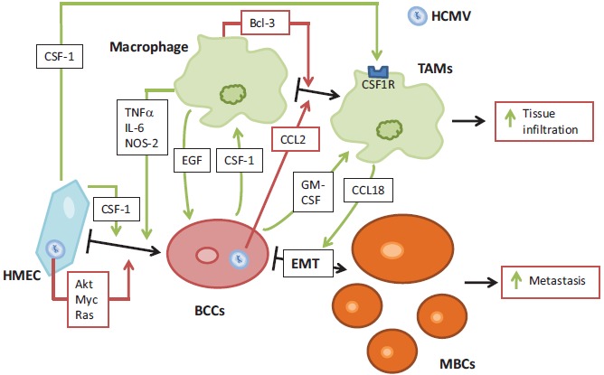
Potential interplay between macrophages, mammary epithelial cells and HCMV in breast cancer. HCMV infection of HMEC induces their priming towards epithelial transformation, by activation of several signaling pathways (Akt, Myc, Ras). HCMV also favors macrophages differentiation towards M2 phenotype. HMEC produces CFS-1, that will enhance cell transformation. This creates a favorable microenvironment, in which infected HMEC produce CCL-2 that promotes TAMs differentiation. In turn, TAMs secrete EGF, that promote breast cancer cells proliferation. Once HMEC had been transformed, they produce GM-CSF, which activate macrophages. In turn, TAMs secrete CCL18, that promotes the epithelial-mesenchymal transition (EMT) of breast cancer cells. This interplay favors cells activations and transformations, leading toward increased metastasis of breast cancer cells and increased tissue infiltration by TAMs.
BCCs: Brest Cancer Cells ; CSF: Colony Stimulating Factor ; EMT: epithelial-mesenchymal transition ; HMEC: Human Mammary Epithelial Cells ; MBCs: Metastatic Breast Cancer cells ; TAMs: Tumor Associated Macrophages ; Bcl: B-cell lymphoma encoded protein ; CCL : C-C Motif Chemokine Ligand ; EGF: Epidermal Growth Factor ; GM: Granulocyte Macrophage ; HCMV : Human Cytomegalovirus ; IL: Interleukin ; NOS: NO Synthase ; TNF: Tumor Necrosis Factor.
Similar articles
-
The oncogenic potential of human cytomegalovirus and breast cancer.Front Oncol. 2014 Aug 25;4:230. doi: 10.3389/fonc.2014.00230. eCollection 2014. Front Oncol. 2014. PMID: 25202681 Free PMC article. Review.
-
A Review of the Potential Role of Human Cytomegalovirus (HCMV) Infections in Breast Cancer Carcinogenesis and Abnormal Immunity.Cancers (Basel). 2019 Nov 22;11(12):1842. doi: 10.3390/cancers11121842. Cancers (Basel). 2019. PMID: 31766600 Free PMC article. Review.
-
The oncomodulatory role of human cytomegalovirus in colorectal cancer: implications for clinical trials.Front Oncol. 2014 Nov 17;4:314. doi: 10.3389/fonc.2014.00314. eCollection 2014. Front Oncol. 2014. PMID: 25452935 Free PMC article. Review.
-
Human Cytomegalovirus Particles Treated with Specific Antibodies Induce Intrinsic and Adaptive but Not Innate Immune Responses.J Virol. 2017 Oct 27;91(22):e00678-17. doi: 10.1128/JVI.00678-17. Print 2017 Nov 15. J Virol. 2017. PMID: 28878085 Free PMC article.
-
Oncomodulatory signals by regulatory proteins encoded by human cytomegalovirus: a novel role for viral infection in tumor progression.FEMS Microbiol Rev. 2004 Feb;28(1):59-77. doi: 10.1016/j.femsre.2003.07.005. FEMS Microbiol Rev. 2004. PMID: 14975530 Review.
Cited by
-
Inflammatory Breast Cancer: The Secretome of HCMV+ Tumor-Associated Macrophages Enhances Proliferation, Invasion, Colony Formation, and Expression of Cancer Stem Cell Markers.Front Oncol. 2022 Jun 30;12:899622. doi: 10.3389/fonc.2022.899622. eCollection 2022. Front Oncol. 2022. PMID: 35847899 Free PMC article.
-
High-Risk Oncogenic Human Cytomegalovirus.Viruses. 2022 Nov 7;14(11):2462. doi: 10.3390/v14112462. Viruses. 2022. PMID: 36366560 Free PMC article. Review.
-
Examining the influence of tumor-infiltrating macrophages on breast cancer outcomes and identifying relevant genes for diagnostic purposes.Discov Oncol. 2024 Sep 27;15(1):502. doi: 10.1007/s12672-024-01397-z. Discov Oncol. 2024. PMID: 39331271 Free PMC article.
-
Identification of UL69 Gene and Protein in Cytomegalovirus-Transformed Human Mammary Epithelial Cells.Front Oncol. 2021 Apr 16;11:627866. doi: 10.3389/fonc.2021.627866. eCollection 2021. Front Oncol. 2021. PMID: 33937031 Free PMC article.
-
Detection of human papillomavirus genotypes, herpes simplex, varicella zoster and cytomegalovirus in breast cancer patients.Virol J. 2021 Jan 22;18(1):25. doi: 10.1186/s12985-021-01498-z. Virol J. 2021. PMID: 33482849 Free PMC article.
References
-
- Canque B., Rosenzwajg M., Gey A., Tartour E., Fridman W.H., Gluckman J.C. Macrophage inflammatory protein-1alpha is induced by human immunodeficiency virus infection of monocyte-derived macrophages. Blood. 1996;87(5):2011–2019. - PubMed
-
- Cotter R.L., Zheng J., Che M., Niemann D., Liu Y., He J., Thomas E., Gendelman H.E. Regulation of human immunodeficiency virus type 1 infection, beta-chemokine production, and CCR5 expression in CD40L-stimulated macrophages: immune control of viral entry. J. Virol. 2001;75(9):4308–4320. doi: 10.1128/JVI.75.9.4308-4320.2001. - DOI - PMC - PubMed
-
- Lisziewicz J., Gabrilovich D.I., Varga G., Xu J., Greenberg P.D., Arya S.K., Bosch M., Behr J.P., Lori F. Induction of potent human immunodeficiency virus type 1-specific T-cell-restricted immunity by genetically modified dendritic cells. J. Virol. 2001;75(16):7621–7628. doi: 10.1128/JVI.75.16.7621-7628.2001. - DOI - PMC - PubMed
Publication types
LinkOut - more resources
Full Text Sources
Other Literature Sources