Case Reports
. 2017 Apr 30;11(4):1-9.
doi: 10.3941/jrcr.v11i4.3010. eCollection 2017 Apr.

Bilateral persistent primitive hypoglossal arteries associated with unilateral symptomatic carotid thromboembolism

Affiliations
Case Reports

Bilateral persistent primitive hypoglossal arteries associated with unilateral symptomatic carotid thromboembolism

Riddhi Patira et al. J Radiol Case Rep. .

Abstract

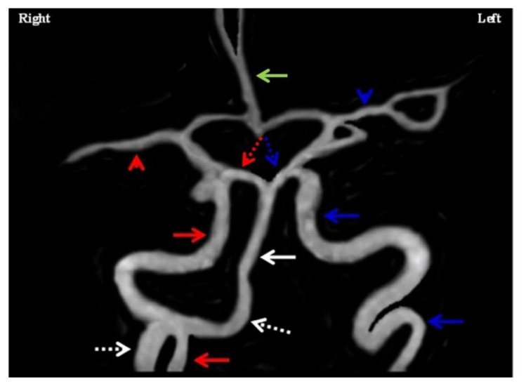
We report the fifth case of bilateral persistent primitive hypoglossal arteries in the literature. This is also the first such case to be demonstrated on computerized tomography angiogram (CTA) and the first case to be associated with a symptomatic carotid thrombus. The sub-occlusive thrombus was distal to the take-off of the dominant persistent hypoglossal artery (PHA) from the internal carotid artery, thus sparing involvement of posterior circulation. Timely identification of the internal carotid artery thromboembolism in the setting of a PHA is important to allow for thrombectomy. Any intervention was not done in this case as the patient was out of the window and at an additional risk of inducing intracranial thromboembolism. Symptomatic carotid stenosis at the PHA take-off is typically treated with endovascular angioplasty and stenting due to the typically high level of the bifurcation in the neck.

Keywords: Bilateral persistent hypoglossal artery; carotid thromboembolism; computed tomography angiogram; endovascular stenting; fetal variant.


Figures

Figure 1
Figure 1
A 79 year-old man with a right middle carotid artery territory infarct secondary to sub-occlusive thrombus within the right internal carotid artery (ICA) in the setting of bilateral persistent hypoglossal arteries (PHAs). Findings: (a) Sagital view of the CTA with vessel-tracking by Tera Recon (white line). The right common carotid artery (inferior aspect of white line depicted by an orange arrow) courses superiorly to branch into the external carotid artery and the ICA. The right ICA continues (white line) until it gives off a right PHA (segment of white line pointed by yellow arrow) and remainder of ICA is further shown to have a sub-occlusive filling defect just distal to the origin of the right PHA, consistent with an intraluminal thrombus (pink arrow). The right PHA (continues as white line pointed by yellow arrow) enters the intracranial space through the right hypoglossal canal (green arrow) and terminates into the basilar artery (blue arrow). (b) Axial view of the CTA and (c) Coronal view of the CTA depicting a large right PHA (red arrow) traversing the hypoglossal canal into the intracranial space, and a smaller left PHA (yellow arrow) entering the left hypoglossal canal. Technique: mA 116, KVp 120, slice thickness 0.60 mm, with 80 mL of Omnipaque 350
Figure 2
Figure 2
A 79 year-old man with a right middle carotid artery territory infarct secondary to sub-occlusive thrombus within the right internal carotid artery (ICA) in the setting of bilateral persistent hypoglossal arteries (PHAs). Findings: Sagital view of CTA with vessel-tracking showing the left PHA (yellow arrow) originating from the left ICA (pink arrow), entering the intracranial space through the left hypoglossal canal (green arrow) to terminate into the ipsilateral posterior inferior cerebellar artery (blue arrow). Technique: mA 116, KVp 120, slice thickness 0.60 mm, with 80 mL of Omnipaque 350
Figure 3
Figure 3
A 79 year-old man with a right middle carotid artery territory infarct secondary to sub-occlusive thrombus within the right internal carotid artery (ICA) in the setting of bilateral persistent hypoglossal arteries (PHAs). Findings: (a) Axial view and (b) Coronal view of CTA depicting a calcified plaque (both red arrows) proximal to the suspected thrombus without any significant stenosis at the level of the plaque. Technique: mA 239, KVp 120, slice thickness 1.00 mm, with 80 mL of Omnipaque 350
Figure 4
Figure 4
A 79 year-old man with a right middle carotid artery territory infarct secondary to sub-occlusive thrombus within the right internal carotid artery (ICA) in the setting of bilateral persistent hypoglossal arteries (PHAs). Findings: (a) Axial diffusion-weighted (DWI) sequence showing a large area of restricted diffusion (white arrow) with (b) matched defect in corresponding ADC map within the territory supplied by the right middle cerebral artery suggesting a right MCA infarct (white arrow). (c) Axial DWI and (d) corresponding ADC map showing cerebellum and brainstem without any areas of restricted diffusion (both white solid arrows). Rest of the areas supplied by the posterior circulation were also spared (not shown here). (e) Axial T2 and (f) coronal proton density sequence showing absence of bilateral vertebral arteries (both red arrows). Technique: (a) – (d): 1.5 T GE Magnet, TR: 9000, TE: 78.8, FOV: 23 cm. (e): 1.5 T GE Magnet, TR: 2516.66, TE: 82.8, FOV: 23 cm. (f): 1.5 T GE Magnet, TR: 2500, TE: 19.66, FOV: 23 cm
Figure 5
Figure 5
A 79 year-old man with a right middle carotid artery territory (MCA) infarct secondary to sub-occlusive thrombus within the right internal carotid artery (ICA) in the setting of bilateral persistent hypoglossal arteries (PHAs). Findings: MRA showing patent basilar artery (white solid arrow) getting its major supply from right PHA (white dotted arrows). Right MCA is patent (red arrowhead). Other patent major intracranial vessels are shown including right ICA (red arrows), left ICA (blue arrows), left MCA (blue arrowhead), right posterior cerebral artery (red dotted arrow), left posterior cerebral artery (blue dotted arrow), and anterior cerebral artery (green solid arrow). Technique: 1.5 T GE Magnet, TR 25, TE: 2.4.
Figure 6
Figure 6
A 79 year-old man with a right middle carotid artery territory infarct secondary to sub-occlusive thrombus within the right internal carotid artery (ICA) in the setting of bilateral persistent hypoglossal arteries (PHAs). Findings: Anatomic illustration showing both PHA (blue segments pointed by blue arrows) originating from respective ICA. In addition, though not present in our case, the location of other fetal variants are shown. The persistent trigeminal artery (white dotted line) is an anastomosis between the distal part of ICA and the distal part of the basilar artery. The persistent otic artery (yellow dotted line) is an anastomosis between the ICA and the proximal part of the basilar artery. The persistent postatlantal artery (green dotted line) is an anastomosis between the proximal part of ICA and the ipsilateral vertebral artery (red dotted line).

Similar articles

Cited by

References

    1. Uchino A, Saito N, Okada Y, Kozawa E, Nishi N, Mizukoshi W, Inoue K, Nakajima R, Takahashi M. Persistent hypoglossal artery and its variants diagnosed by CT and MR angiography. Neuroradiology. 2013 Jan 1;55(1):17–23. - PubMed
    1. Karasawa J, Kikuchi HA, Furuse SE, Sakaki TO, Yoshida YA, Ohnishi HI. Bilateral persistent carotid-basilar anastomoses. American Journal of Roentgenology. 1976 Dec 1;127(6):1053–6. - PubMed
    1. Murayama Y, Fujimoto N, Matsumoto K. Bilateral persistent primitive hypoglossal arteries associated with a large ruptured aneurysm on one side. Surgical neurology. 1985 Nov 30;24(5):498–502. - PubMed
    1. Garge S, Moses V, Keshava S, Ahmed M, Moorthy R. Persistent hypoglossal arteries with aneurysmal dilation of left hypoglossal artery: a rare case report and review of the literature. BJR case reports. 2016 May;:20150301. - PMC - PubMed
    1. Takahashi H, Tanaka H, Fujita N, Tomiyama N. Bilateral persistent hypoglossal arteries: MRI findings. The British journal of radiology. 2014 Jan 28; - PMC - PubMed

Publication types

LinkOut - more resources