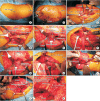
Rotational Acetabular Osteotomy
- PMID: 28567213
- PMCID: PMC5435649
- DOI: 10.4055/cios.2017.9.2.129
Rotational Acetabular Osteotomy
Abstract
Hip dysplasia is the most common cause of secondary osteoarthritis (OA). To prevent the early onset of secondary OA, Nishio's transposition osteotomy, Steel's triple osteotomy, Eppright's dial osteotomy, Wagner's spherical acetabular osteotomy, Tagawa's rotational acetabular osteotomy (RAO), and Ganz' periacetabular osteotomy (PAO) have been proposed. PAO and RAO are now commonly used in surgical treatment of symptomatic acetabular dysplasia in Europe, North America, and Asia. The aim of this paper is to present the followings: the patient selection criteria for RAO; the surgical technique of RAO; the long-term outcome of RAO; and the future perspectives.
Keywords: Hip dysplasia; Pelvic osteotomy.
Conflict of interest statement
CONFLICT OF INTEREST: No potential conflict of interest relevant to this article was reported.
Figures


References
-
- Aronson J. Osteoarthritis of the young adult hip: etiology and treatment. Instr Course Lect. 1986;35:119–128. - PubMed
-
- Harris WH. Etiology of osteoarthritis of the hip. Clin Orthop Relat Res. 1986;(213):20–33. - PubMed
-
- Murphy SB, Ganz R, Muller ME. The prognosis in untreated dysplasia of the hip: a study of radiographic factors that predict the outcome. J Bone Joint Surg Am. 1995;77(7):985–989. - PubMed
-
- Nishio A. Transposition osteotomy of the acetabulum for the treatment of congenital dislocation of the hip. Nippon Seikeigeka Gakkai Zasshi. 1956;30:482–484.
-
- Steel HH. Triple osteotomy of the innominate bone. J Bone Joint Surg Am. 1973;55(2):343–350. - PubMed
Publication types
MeSH terms
LinkOut - more resources
Full Text Sources
Other Literature Sources
Medical

