

Circulatory zinc transport is controlled by distinct interdomain sites on mammalian albumins
- PMID: 28567254
- PMCID: PMC5450522
- DOI: 10.1039/c6sc02267g
Circulatory zinc transport is controlled by distinct interdomain sites on mammalian albumins
Abstract
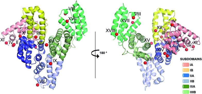
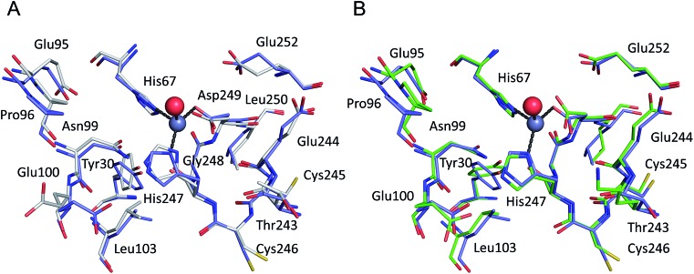
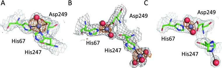
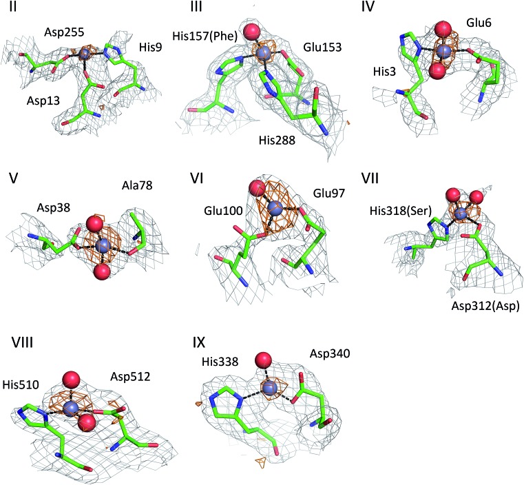
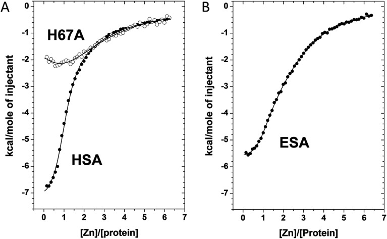
Zinc is an essential nutrient in the body; it is required for the catalytic activity of many hundreds of human enzymes and virtually all biological processes, therefore its homeostasis and trafficking is of crucial interest. Serum albumin is the major carrier of Zn2+ in the blood and is required for its systemic distribution. Here we present the first crystal structures of human serum albumin (HSA) and equine serum albumin (ESA) in complex with Zn2+. The structures allow unambiguous identification of the major zinc binding site on these two albumins, as well as several further, weaker zinc binding sites. The major site in both HSA and ESA has tetrahedral geometry and comprises three protein ligands from the sidechains of His67, His247 and Asp249 and a water molecule. Isothermal titration calorimetric studies of a HSA H67A mutant confirm this to be the highest affinity Zn2+ site. Furthermore, analysis of Zn2+ binding to HSA and ESA proved the presence of secondary sites with 20-50-fold weaker affinities, which may become of importance under particular physiological conditions. Both calorimetry and crystallography suggest that ESA possesses an additional site compared to HSA, involving Glu153, His157 and His288. The His157 residue is replaced by Phe in HSA, incapable of metal coordination. Collectively, these findings are critical to our understanding of the role serum albumin plays in circulatory Zn2+ handling and cellular delivery.
Figures
References
-
- Peters T. J. Clin. Chem. 1977;23:5–12. - PubMed
-
- Fanali G., di Masi A., Trezza V., Marino M., Fasano M., Ascenzi P. Mol. Aspects Med. 2012;33:209–290. - PubMed
-
- He X. M., Carter D. C. Nature. 1992;358:209–215. - PubMed
-
- Bal W., Sokołowska M., Kurowska E., Faller P. Biochim. Biophys. Acta, Gen. Subj. 2013;1830:5444–5455. - PubMed
-
- Giroux E. L., Henkin R. I. Biochim. Biophys. Acta, Gen. Subj. 1972;273:64–72. - PubMed
Grants and funding
LinkOut - more resources
Full Text Sources
Other Literature Sources
Molecular Biology Databases