

Successful percutaneous closure of an extremely large secundum atrial septal defect during pregnancy
- PMID: 28567360
- PMCID: PMC5440271
- DOI: 10.21037/cdt.2016.10.03
Successful percutaneous closure of an extremely large secundum atrial septal defect during pregnancy
Abstract
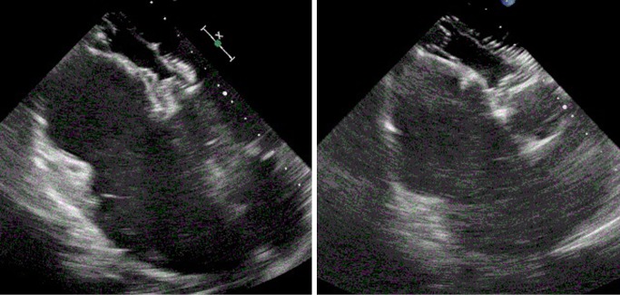
Atrial septal defects (ASDs) are one of the most of the most common acyanotic congenital heart lesions. Awareness of potential clinical presentations and complications during pregnancy is essential for those managing these patients. We report successful percutaneous closure of an extremely large secundum ASD, using the largest available percutaneous ASD closure device in a 27-year-old pregnant female. Large ASDs may have their initial clinical presentation and diagnosis during pregnancy. If indicated, percutaneous closure can be performed safely. Only a very small number of cases have previously reported this being performed safely during pregnancy.
Keywords: Atrial septal defect (ASD); percutaneous closure; pregnancy.
Conflict of interest statement
Conflicts of Interest: The authors have no conflicts of interest to declare.
Figures

Similar articles
-
Clinical results of large secundum atrial septal defect closure in adult using percutaneous transcatheter Cocoon atrial septal occluder.J Med Assoc Thai. 2013 Sep;96(9):1127-34. J Med Assoc Thai. 2013. PMID: 24163987
-
Transcatheter closure of secundum atrial septal defects: results in patients with large and extreme defects.Heart Lung Circ. 2014 Feb;23(2):127-31. doi: 10.1016/j.hlc.2013.07.020. Epub 2013 Sep 4. Heart Lung Circ. 2014. PMID: 24012104
-
[Percutaneous transcatheter atrial septal defect closure with Amplatzer septal occluder device using three different techniques in three adult patients with complex ostium secundum type atrial defects].Turk Kardiyol Dern Ars. 2013 Mar;41(2):148-53. doi: 10.5543/tkda.2013.79745. Turk Kardiyol Dern Ars. 2013. PMID: 23666304 Turkish.
-
Recent advances in managing septal defects: atrial septal defects.F1000Res. 2017 Nov 22;6:2042. doi: 10.12688/f1000research.11844.1. eCollection 2017. F1000Res. 2017. PMID: 29250321 Free PMC article. Review.
-
Interventional Therapy Versus Medical Therapy for Secundum Atrial Septal Defect: A Systematic Review (Part 2) for the 2018 AHA/ACC Guideline for the Management of Adults With Congenital Heart Disease: A Report of the American College of Cardiology/American Heart Association Task Force on Clinical Practice Guidelines.J Am Coll Cardiol. 2019 Apr 2;73(12):1579-1595. doi: 10.1016/j.jacc.2018.08.1032. Epub 2018 Aug 16. J Am Coll Cardiol. 2019. PMID: 30121241
Cited by
-
Transoesophageal echocardiography-guided balloon-assisted percutaneous closure of a large secundum atrial septal defect in a pregnant woman: a case report.Eur Heart J Case Rep. 2024 Jan 6;8(1):ytae014. doi: 10.1093/ehjcr/ytae014. eCollection 2024 Jan. Eur Heart J Case Rep. 2024. PMID: 38274706 Free PMC article.
References
-
- INTECH. Pregnancy Issues in Women with Atrial Septal Defect. Available online: http://www.intechopen.com/books/atrial-septal-defect/pregnancy-issues-in...
Publication types
LinkOut - more resources
Full Text Sources
Other Literature Sources