

Coeliac disease and the videocapsule: what have we learned till now
- PMID: 28567377
- PMCID: PMC5438789
- DOI: 10.21037/atm.2017.05.06
Coeliac disease and the videocapsule: what have we learned till now
Abstract
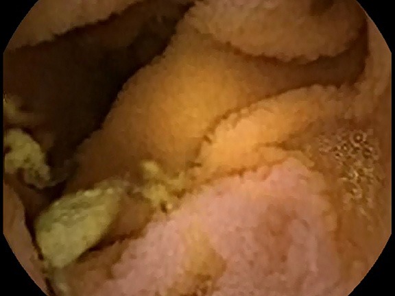
Celiac disease is diagnosed in part by finding areas of pathology in the small bowel (SB) mucosa. This can often be difficult because the pathologic alterations, including atrophy of the small intestinal villi, can often be sparse and subtle. Some of the quantitative methods for detecting and measuring the presence of villous atrophy from videocapsule endoscopy (VCE) images are presented and discussed. These methods consist of static features of measurement including texture, gray level, and presence and abundance of fissures contained within each acquired image. The methods also consist of dynamic measurements including spectral analysis, and determining motion from a sequence of endoscopic images as obtained from a VCE clip. Thus far, several methods have been found useful to characterize the SB mucosa of untreated celiac disease patients versus control patients lacking villous atrophy, which have revealed significant differences in texture, frequency, and motion on analysis of VCE. In untreated celiac patients undergoing endoscopy, there tends to be greater magnitude of changes and spatial differences in textural descriptors, longer periodic components, indicating slower periodic activity, and differences in feature location, suggesting alterations in motility at areas of pathology as compared to patients without villous atrophy. Improvements in the quantitative analysis of VCE imaging in celiac patients is important to detect pathology in suspected patients, so that biopsies can be obtained from pertinent regions of the small intestinal mucosa. Improvements are also necessary so that patients with celiac disease can be monitored to evaluate the progress of mucosal healing after onset of treatment.
Keywords: Celiac disease; endoscopy; small intestine; videocapsule; villous atrophy.
Conflict of interest statement
Conflicts of Interest: The authors have no conflicts of interest to declare.
Figures


Similar articles
-
Extraction and processing of videocapsule data to detect and measure the presence of villous atrophy in celiac disease patients.Comput Biol Med. 2016 Nov 1;78:97-106. doi: 10.1016/j.compbiomed.2016.09.009. Epub 2016 Sep 16. Comput Biol Med. 2016. PMID: 27673492
-
Methods to quantitate videocapsule endoscopy images in celiac disease.Biomed Mater Eng. 2014;24(6):1895-911. doi: 10.3233/BME-140999. Biomed Mater Eng. 2014. PMID: 25226886 Review.
-
Transformation of videocapsule images to detect small bowel mucosal differences in celiac versus control patients.Comput Methods Programs Biomed. 2012 Oct;108(1):28-37. doi: 10.1016/j.cmpb.2011.12.008. Epub 2012 Jan 28. Comput Methods Programs Biomed. 2012. PMID: 22284703
-
Implementation of a polling protocol for predicting celiac disease in videocapsule analysis.World J Gastrointest Endosc. 2013 Jul 16;5(7):313-22. doi: 10.4253/wjge.v5.i7.313. World J Gastrointest Endosc. 2013. PMID: 23858375 Free PMC article.
-
Suggestions for automatic quantitation of endoscopic image analysis to improve detection of small intestinal pathology in celiac disease patients.Comput Biol Med. 2015 Oct 1;65:364-8. doi: 10.1016/j.compbiomed.2015.04.019. Epub 2015 Apr 24. Comput Biol Med. 2015. PMID: 25976612 Review.
Cited by
-
Current Evidence on Computer-Aided Diagnosis of Celiac Disease: Systematic Review.Front Pharmacol. 2020 Apr 16;11:341. doi: 10.3389/fphar.2020.00341. eCollection 2020. Front Pharmacol. 2020. PMID: 32372947 Free PMC article.
-
Automated diagnosis of celiac disease by video capsule endoscopy using DAISY Descriptors.J Med Syst. 2019 Apr 26;43(6):157. doi: 10.1007/s10916-019-1285-6. J Med Syst. 2019. PMID: 31028562
References
Publication types
LinkOut - more resources
Full Text Sources
Other Literature Sources