. 2017 Jun 1;8(6):e2836.
doi: 10.1038/cddis.2017.67.

Interferon-γ derived from cytotoxic lymphocytes directly enhances their motility and cytotoxicity

Affiliations

Interferon-γ derived from cytotoxic lymphocytes directly enhances their motility and cytotoxicity

Purnima Bhat et al. Cell Death Dis. .

Abstract

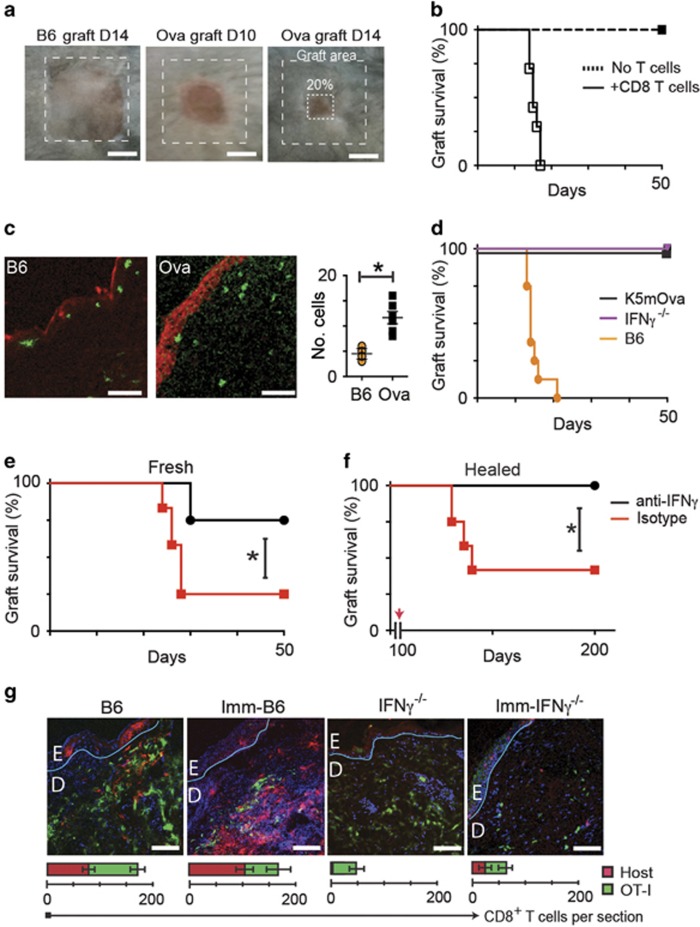
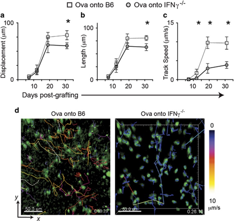
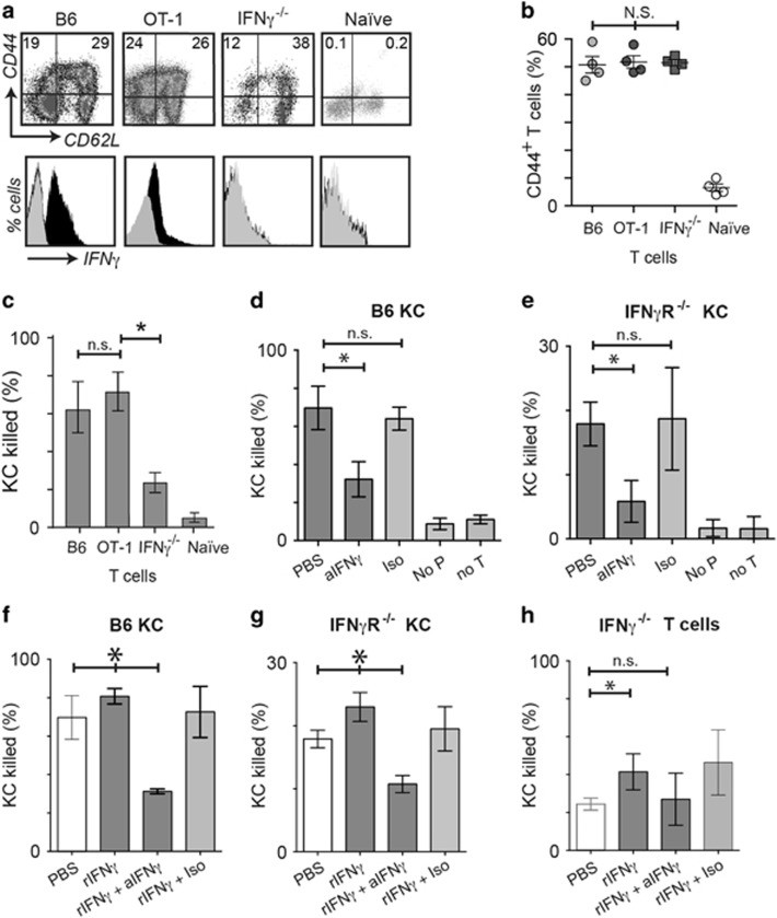
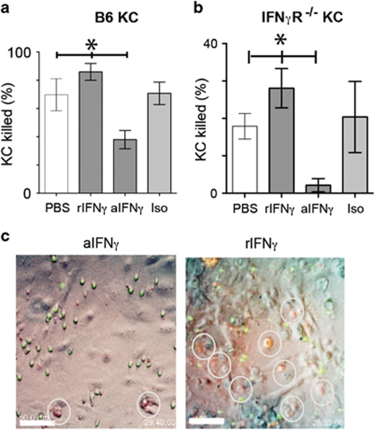
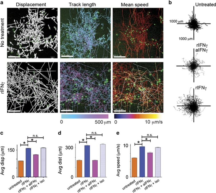
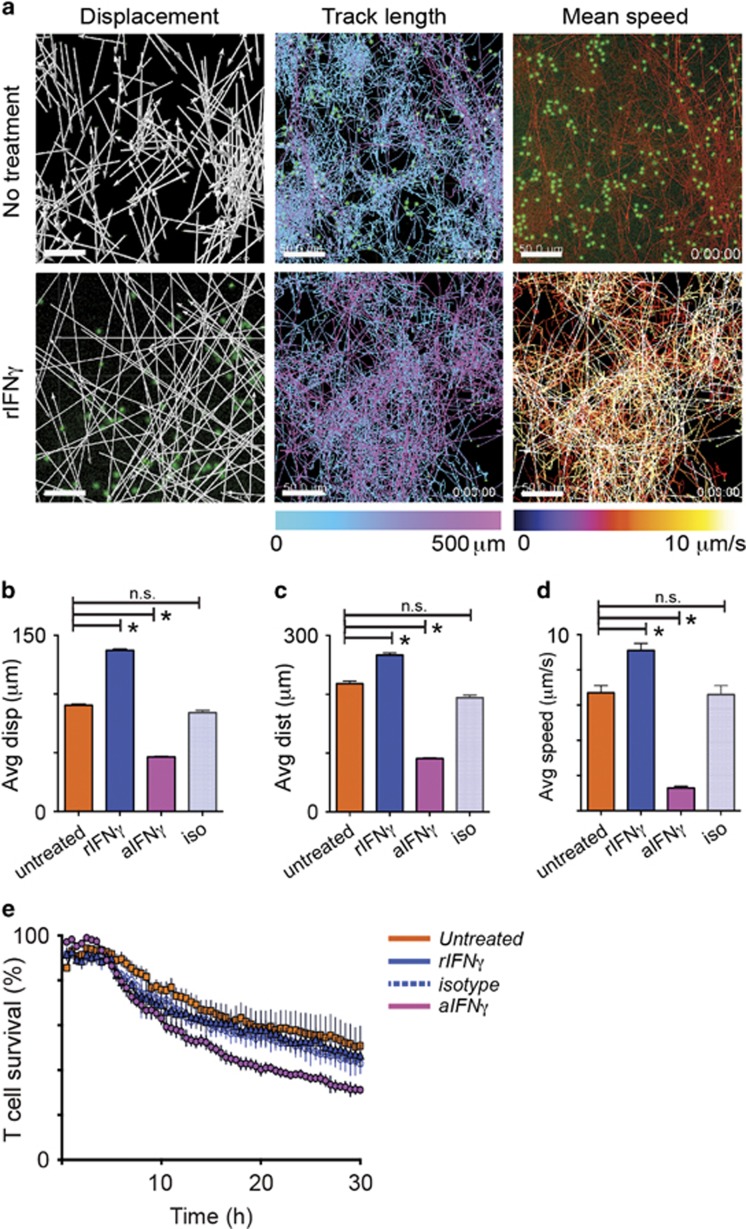
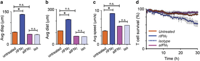
Interferon gamma (IFNγ) is a key moderator of cell-mediated immunity with diverse, mainly pro-inflammatory actions on immunocytes and target tissue. Recent studies have shown it may enhance anti-tumor and antiviral effects of CD8 T cells. Here we investigate the mechanisms by which IFNγ mediates CD8 T-cell cytotoxic function. We show that in vivo, antigen-specific CD8 T cells that produce INFγ are necessary to effect rejection of skin grafts expressing OVA as a transgene in keratinocytes. The ability of CD8 T cells to produce IFNγ enhanced their ability to migrate to the site of antigen-presenting skin cells. By in vivo imaging, we show that CTL motility, particularly speed, during graft rejection was enhanced by locally available IFNγ. We then used a reductionist two-cell model of CTL effectors and keratinocyte targets to investigate the effects of locally available (paracrine) and CTL-producing (autocrine) IFNγ on the motility behavior and killing ability of the CTL. Using live-cell imaging by prolonged time-lapse microscopy of primary effector CD8 T cells and antigen-expressing primary keratinocyte targets, we show that CD8 T-cell cytotoxic function and motility is enhanced by locally available IFNγ. Conversely, deprivation of either autocrine or paracrine IFNγ, or blockade of IFNγ signaling to CTL markedly reduced their cytotoxic function, their kinematics, and effector cell survival. We conclude that in vitro and in vivo, autocrine production of IFNγ by CTL enhances their motility and promotes killing of primary target keratinocytes. The absolute need for local IFNγ to enable cytotoxic CD8 T-cell function is of significance for immunotherapy for chronic viral infection and for cancer.


Conflict of interest statement

The authors declare no conflict of interest.

Figures

Figure 1
Figure 1
Interferon-γ is required for skin graft rejection. Ear skin from B6 or K5mOVA donor mice was grafted on the flanks of B6 recipients. (a) 80% graft loss was denoted as rejection. (b) OVA skin grafted onto Rag1−/− mice with or without transferred 106 naive CD8 T cells. (c) Section of OVA grafts onto B6 or OVA mice at day 10 stained for caspase-3 (red), CD8 (green; Bar, 100 μm); quantified per field of view. Data pooled from 2 experiments, error bars are SEM. (d) OVA transgenic grafts were placed on K5mOva, IFNγ−/− or B6 recipients. Graph shows graft survival. (e) IFNγ−/− recipients received pre-primed CD8 cells by adoptive transfer, and were treated with anti-IFNγ or isotype antibody 48 h prior to grafting of OVA skin, and weekly thereafter. Graph shows graft survival (*P<0.05, Mantel–Cox test). (f) OVA skin grafts were placed on IFNγ−/− mice for 100 days. Recipients were then adoptively transferred with IFNγ-competent CTL, and anti-IFNγ or isotype antibody as in (e). (*P<0.001, Mantel–Cox test). (g) EGFP+ OT-1 CD8 T cells were adoptively transferred into OVA-naive B6 mice, OVA-immunized B6 mice, OVA-naive IFNγ−/− mice, or into OVA-immunized IFNγ−/− mice, before placing OVA skin grafts. Grafts were harvested after 10 days and stained for CD8. Confocal images were taken at graft edges and have been pseudocoloured to show native CD8 cells (red), transferred cells (green) and nuclei (blue). The basement membranes have been drawn in teal. (Bar, 100 μm). The average total cell numbers of T cells in each field from duplicate fields of two samples from a typical experiment is indicated in the graphs below. Error bars are S.D.
Figure 2
Figure 2
Motility of CTL in tissues is enhanced by IFNγ. EGFP+CTL were adoptively transferred into B6 or IFNγ−/− mice before OVA skin was grafted. Post grafting, mice were anaesthetized and the graft edges imaged by 2-photon microscopy. Multiple sites per graft were imaged for 20-30 min each. EGFP+ cells were identified and tracked by software, and analyzed for CTL displacement (a), length of travel (b) and speed of travel (c) during the imaged time. Data are pooled from 4 regions imaged per graft, from 3 mice per time point. Averages are shown, error bars are SD. (*P<0.05) (d) Representative images of grafts on B6 recipients and IFNγ−/− recipients at day 19 post graft placement. CTL have been identified and tracked with track color coding for average speed of cell movement over the period of imaging. Bar, 50 μm (see also Supplementary Movies S1 and 2)
Figure 3
Figure 3
Exogenous IFNγ promotes KC killing by T cells via IFNγ receptor on target cells. (a) Flow cytometry analysis of splenic T cells gated by size, CD8 expression, and EGFP expression, from OVA-immunized OT-I, B6, IFNγ−/−, or OVA-naive B6 mice (black, anti-IFNγ; gray, isotype antibody). N=4. (N.S., not significant between groups, ANOVA.) (b) Percentage of total CD8 T cells from (a) that express CD44. N=4, error bars are SD. (ch) These cells were co-cultured with SIINFEKL-loaded target keratinocytes derived from B6 (IFNγR+/+) or IFNγR−/− mice in the presence of indicator dye for intracellular activated caspases, and imaged by time-lapse fluorescence microscopy. Target cell death was determined by analyzing movies using software to identify spots based on size (>12 μm) and red fluorescence, and expressed as a percentage of the total number of KC at 30 h. Duplicate wells were averaged, N=4. Error bars are standard deviations. (*P<0.05, ‘NS’, not significant, Mann–Whitney test; *P<0.05, ‘n.s.’, not significant, ANOVA) (d) OT-I CTL were co-cultured with IFNγR+/+ or IFNγR−/− (e) KC. Co-cultures were treated with anti-IFNγ or PBS. Included in each experiment were control wells: with isotype antibody (Iso), without peptide (No P), and without T cells (No T). (f) OT-I CTL were co-cultured with IFNγR+/+ or IFNγR−/− (g) KC and were treated with rIFNγ, or rIFNγ and anti-IFNγ, or rIFNγ and isotype antibody. (h) IFNγ−/− CD8 T cells were co-cultured with SIINFEKL-loaded B6 KC. Cultures were treated with rIFNγ, or rIFNγ and anti-IFNγ, or rIFNγ and isotype antibody
Figure 4
Figure 4
Modulation of IFNγ signaling in CTL affects cytotoxic function. EGFP+OT-1 CTL were incubated with PBS, rIFNγ, anti-IFNγ or isotype antibody for 4 h, then washed thoroughly and added to SIINFEKL-loaded (a) B6 or (b) IFNγR−/− KC along with indicator dye for activated caspase. Co-cultures proceeded for 30 h, and were imaged by fluorescence time-lapse microscopy. Keratinocyte death was determined as in Figure 3. (*P<0.05, ‘NS’, not significant, ANOVA). (c) Frames of representative fields of co-cultures after 28 h of imaging. Merged red, green and brightfield images shown. Dead KC fluoresce red. EGFP+ CTL are green. Dead KC are shown by white rings. Scale bar is 50 μm
Figure 5
Figure 5
Exogenous IFNγ promotes CD8 T-cell motility and enhances cytotoxicity. (a) Co-cultures of EGFP+OT-I CTL and SIINFEKL-loaded or untreated KC were imaged for 30 h by fluorescence time-lapse microscopy. Recombinant IFNγ was added at time 0. Representative images of tracks created by CTL as they traveled among KC targets have been mapped by software. Vector displacement of the tracks is shown by white arrows. Length of the traveled tracks and mean speed of travel is indicated by the colored legend bars (scale bars, 50 μm). (b) Translated travel tracks from (a) have been plotted from origin showing the distance and spread of the path traveled by the cells within the field of view. Each plot shows the result for one typical experiment of three independent experiments. (ce) Displacement (c), distance traveled (d) and speed (e) of CD8 T cells in co-culture with antigen-loaded target KC. Average values of four movies are shown (>400 cells). Error bars are S.E.M. (*P<0.05, n.s. not significant)
Figure 6
Figure 6
Suppression of autocrine signaling of IFNγ on CTL inhibits their cytotoxicity and motility. IFNγ−/− CTL cells or EGFP+OT-I CTL pre-treated with PBS, rIFNγ or anti-IFNγ for 4 h, were co-cultured with SIINFEKL-loaded target KC. The kinematics of CTL were analyzed using fluorescence time-lapse movies of 30 h as above. (a) Examples of images of tracked CD8 T cells. Displacement is shown in white arrows, and track length and mean speed are shown in color. Scale bars, 50 μm. (bd) Pre-treated B6 CTL or IFNγ−/− CTL in co-culture with targets for 30 h were investigated for vectorial displacement, distance traveled, and mean speed. Data are pooled results of 3 independent experiments (around 200 cells). Error bars are SEM. (*P<0.05, n.s. not significant) (e) Rate of death of pre-treated CD8 T cells during co-culture with target cells. The number of surviving T cells was determined by software, expressing the number of green cells (size >7 μm) at each frame as a percentage of the number of cells at the start. N=3. Error bars are s.e.m.
Figure 7
Figure 7
IFNγ-deficient cells show reduced kinematics but respond to IFNγ. CTL from OVA-immunized IFNγ-/- mice were pre-treated with rIFNγ or aIFNγ or isotype control antibody for four hours prior to co-culture with KC presenting SIINFEKL peptide. Cultures were imaged for 30 h as above. (a-c) Movies were analyzed for CTL displacement, track length, and average track speed. (d) CTL survival was calculated by comparing the number of CTL in the final imaging frames with those in the initial frames. (*P<0.05, n.s., not significant)

Similar articles

Cited by

References

    1. Martinet L, Garrido I, Filleron T, Le Guellec S, Bellard E, Fournie JJ et al. Human solid tumors contain high endothelial venules: association with T- and B-lymphocyte infiltration and favorable prognosis in breast cancer. Cancer Res 2011; 71: 5678–5687. - PubMed
    1. Peng LS, Zhuang Y, Shi Y, Zhao YL, Wang TT, Chen N et al. Increased tumor-infiltrating CD8(+)Foxp3 (+) T lymphocytes are associated with tumor progression in human gastric cancer. Cancer Immunol Immunother 2012; 61: 2183–2192. - PMC - PubMed
    1. Bellavance EC, Kohlhapp FJ, Zloza A, O'Sullivan JA, McCracken J, Jagoda MC et al. Development of tumor-infiltrating CD8+ T cell memory precursor effector cells and antimelanoma memory responses are the result of vaccination and TGF-β blockade during the perioperative period of tumor resection. J Immunol 2011; 186: 3309–3316. - PMC - PubMed
    1. Sikora AG, Jaffarzad N, Hailemichael Y, Gelbard A, Stonier SW, Schluns KS et al. IFN-α enhances peptide vaccine-induced CD8+ T cell numbers, effector function, and antitumor activity. J Immunol 2009; 182: 7398–7407. - PMC - PubMed
    1. Ruocco MG, Pilones KA, Kawashima N, Cammer M, Huang J, Babb JS et al. Suppressing T cell motility induced by anti-CTLA-4 monotherapy improves antitumor effects. J Clin Invest 2012; 122: 3718–3730. - PMC - PubMed

MeSH terms

Substances