. 2017 Jun 1;8(6):e2845.
doi: 10.1038/cddis.2017.234.

Disease-linked connexin26 S17F promotes volar skin abnormalities and mild wound healing defects in mice

Affiliations

Disease-linked connexin26 S17F promotes volar skin abnormalities and mild wound healing defects in mice

Eric Press et al. Cell Death Dis. .

Erratum in

Abstract

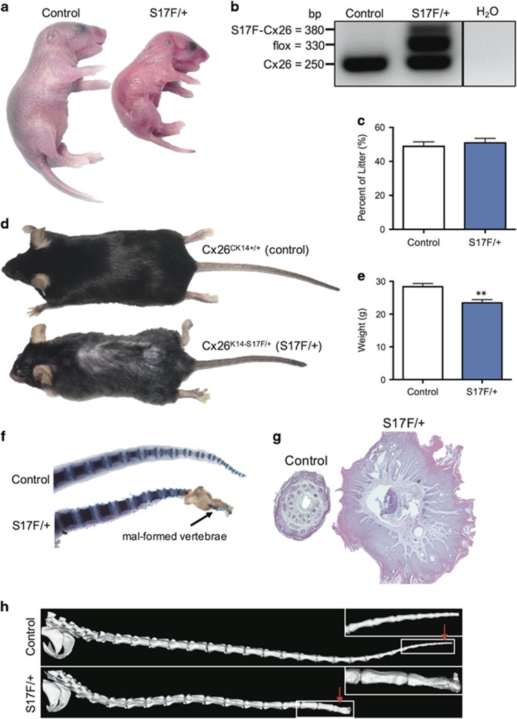
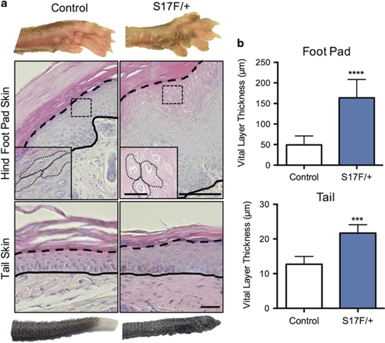
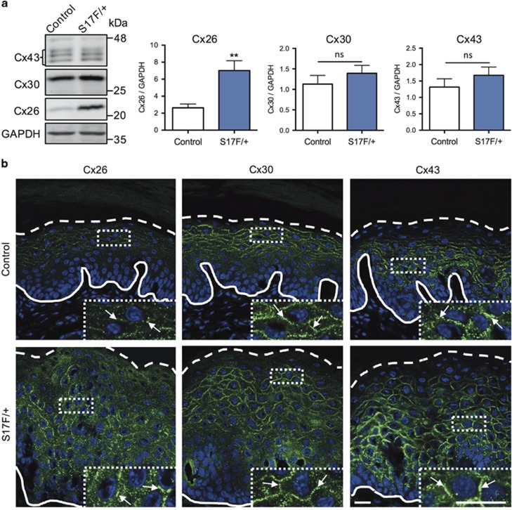
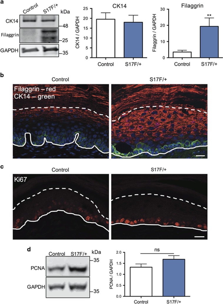
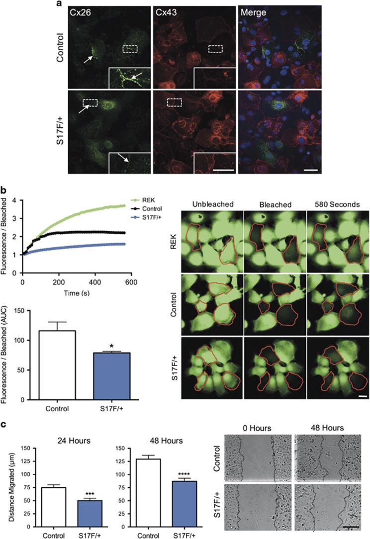
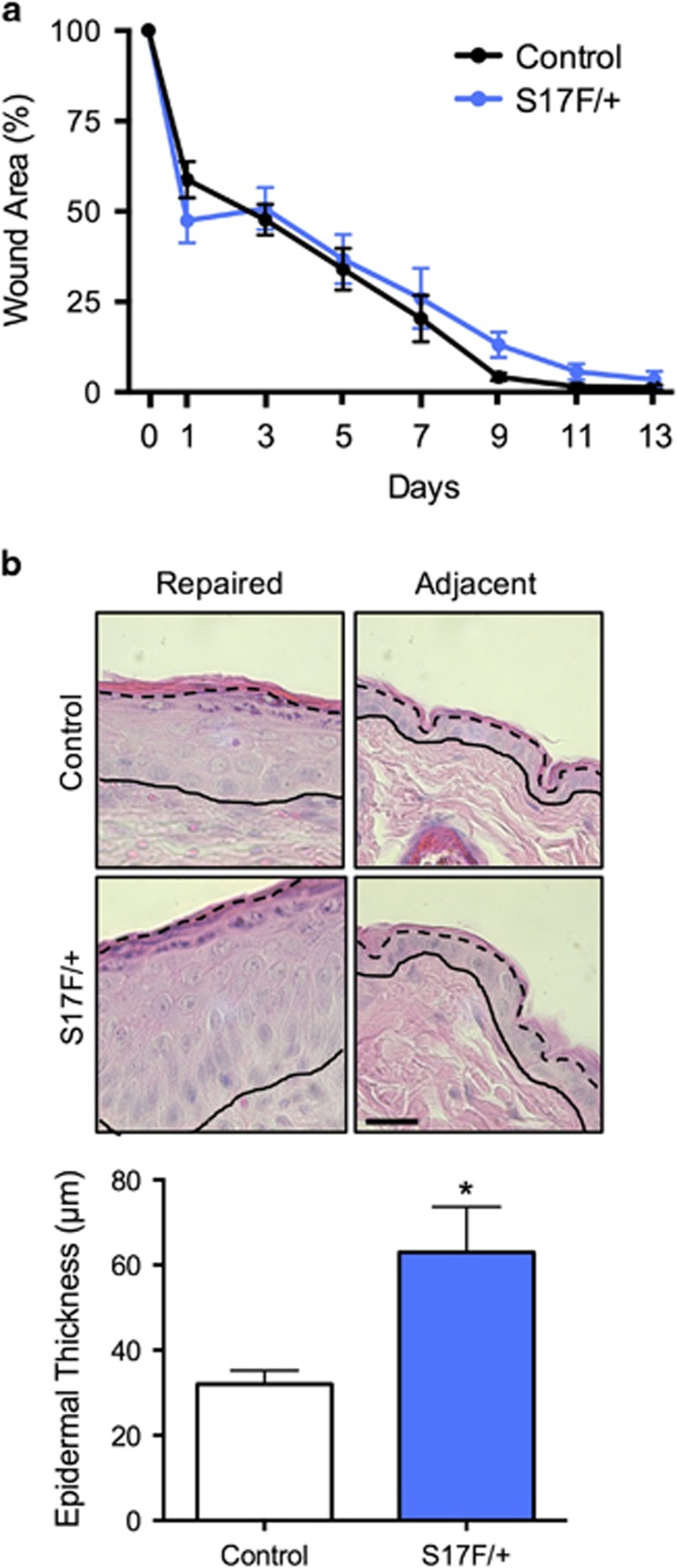
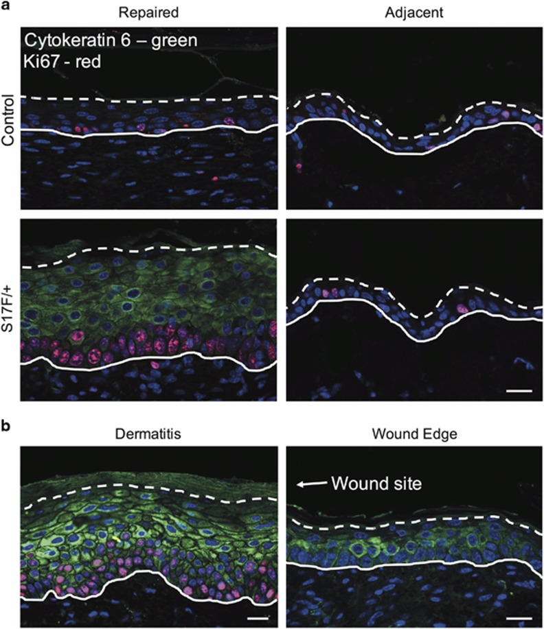
Several mutant mice have been generated to model connexin (Cx)-linked skin diseases; however, the role of connexins in skin maintenance and during wound healing remains to be fully elucidated. Here we generated a novel, viable, and fertile mouse (Cx26CK14-S17F/+) with the keratitis-ichthyosis-deafness mutant (Cx26S17F) driven by the cytokeratin 14 promoter. This mutant mouse mirrors several Cx26-linked human skin pathologies suggesting that the etiology of Cx26-linked skin disease indeed stems from epidermal expression of the Cx26 mutant. Cx26CK14-S17F/+ foot pad epidermis formed severe palmoplantar keratoderma, which expressed elevated levels of Cx26 and filaggrin. Primary keratinocytes isolated from Cx26CK14-S17F/+ neonates exhibited reduced gap junctional intercellular communication and migration. Furthermore, Cx26CK14-S17F/+ mouse skin wound closure was normal but repaired epidermis appeared hyperplastic with elevated expression of cytokeratin 6. Taken together, we suggest that the Cx26S17F mutant disturbs keratinocyte differentiation and epidermal remodeling following wound closure. We further posit that Cx26 contributes to epidermal homeostasis by regulating keratinocyte differentiation, and that mice harboring a disease-linked Cx26 mutant display epidermal abnormalities yet retain most wound healing properties.


Conflict of interest statement

The authors declare no conflict of interest.

Figures

Figure 1
Figure 1
S17F/+ mice mimic KIDS skin characteristics and have several additional phenotypes. (a) S17F/+ neonates were smaller and visually distinguishable from control littermates. (b) Skin sample PCR amplification confirms the heterozygous expression of Cx26S17F in S17F/+ mice. (c) Litters contained equal portions of S17F/+ and control pups with no loss of mouse viability (unpaired t-test, Control: n=157, S17F/+: n=148). (d) Representative photos of 3-month-old S17F/+ and control mice reveal moderate differences in size but a pronounced tail phenotype. (e) Whole mouse weights at 3 months of age showed S17F/+ mice were ~15% smaller (unpaired t-test, **P<0.01, N=17). (f) Skeletal stains of P7 tails revealed vertebral malformations underlying the tail phenotype in S17F/+ mice. (g) Cross-section of a distal tail paraffin section stained with hematoxylin and eosin revealed grossly thickened epidermis. (h) μCT scans of 3-month-old mouse tails revealed vertebral abnormalities in S17F/+ mice. Red arrows in (h) denote the relative locations of cross sections in (g)
Figure 2
Figure 2
S17F/+ neonates display an intact epidermal barrier. P2 neonates were submerged in an aqueous toluidine blue solution to assess epidermal barrier permeability. Similar to control littermates, S17F/+ neonates displayed no epidermal staining indicating a functional barrier (a). To demonstrate epidermal staining from a defective barrier, neonatal epidermis was cut or treated with acetone (b). N=6
Figure 3
Figure 3
S17F/+ mice display thicker foot pad and tail epidermis. (a) 3-month-old S17F/+ mice exhibit severe foot pad epidermal thickening including abnormal non-squamous keratinocytes in suprabasal layers (insets) as well as thicker tail epidermis compared to controls (b) (unpaired t-test, ***P<0.001, ****P<0.0001, N=7). Complete and dashed lines denote the dermis-epidermis boundary and stratum granulosum-corneum boundary, respectively. Scale bar in (a) (upper), 50 μm (inset=10 μm); (a) (lower), 10 μm
Figure 4
Figure 4
S17F/+ foot pad skin exhibits deregulated connexin expression. (a) Foot pad skin lysates from 3-month-old S17F/+ mice exhibited elevated levels of Cx26 expression compared to controls (unpaired t-test, **P<0.01, ns=P>0.05, N=10). (b) Cryosections of 3-month-old S17F/+ foot pad epidermis revealed that Cx26, Cx30, and Cx43 formed abundant gap junctions in a broad range of keratinocyte layers. Complete and dashed lines denote the dermis-epidermis boundary and stratum granulosum-corneum boundary, respectively. Scale bar, 20 μm (inset=20 μm)
Figure 5
Figure 5
S17F/+ mice display abnormal keratinocyte differentiation in foot pad epidermis. (a) Foot pad skin lysates from 3-month-old S17F/+ mice have elevated filaggrin, but normal keratin 14 levels compared with controls (unpaired t-test, **P<0.01, N=10). (b) 3-month-old foot pad epidermis labeled for filaggrin (red) and cytokeratin 14 (green) revealed elevated filaggrin labeling of suprabasal keratinocytes in S17F/+ epidermis. (c) 3-month-old foot pad epidermis labeled for Ki67 (red) and lysates immunoblotted for PCNA (d) demonstrated unaltered levels of cell proliferation in S17F/+ epidermis (unpaired t-test, ns=P>0.05, N=10). Complete and dashed lines denote the dermis-epidermis boundary and stratum granulosum-corneum boundary, respectively. Scale bar in (b and c)=20 μm
Figure 6
Figure 6
Keratinocytes isolated from S17F/+ neonates have reduced GJIC and collective cell migration. (a) S17F/+ keratinocyte cultures appeared to form fewer and smaller Cx26 gap junctions between cells. (b) S17F/+ keratinocytes have reduced calcein-AM fluorescence recovery after photobleaching compared to controls indicative of reduced GJIC (photobleached cells are outlined in red) (unpaired t-test, *P<0.05, N=3). (c) Collective keratinocyte migration in response to scratch wounds was reduced in S17F/+ cultures compared to controls (unpaired t-test, ***P<0.001, ****P<0.0001, N=4). Scale bar in (a)=20 μm (inset=10 μm), (b)=10 μm, (c)=100 μm
Figure 7
Figure 7
S17F/+ mice display normal wound closure, but exhibit abnormal epidermis remodeling. (a) Dorsal skin wound closure was found to be similar between S17F/+ mice and controls (c) (two-way ANOVA with repeated measures), however, epidermis remodeling following wound closure generated thicker epidermis in S17F/+ mice compared with controls (b) (unpaired t-test, *P<0.05, N=4). Scale bar, 10 μm
Figure 8
Figure 8
Repaired S17F/+ epidermis displays activated keratinocytes 14 days following wounding. (a) Repaired S17F/+ epidermis revealed prominent expression of cytokeratin 6 (green) and Ki67 (red) indicating activated keratinocytes similar to skin exhibiting dermatitis (b). Cytokeratin 6 expression diminishes at the wound edge (b). Scale bar in (a and b)=20 μm

Similar articles

Cited by

References

    1. Alexander DB, Goldberg GS. Transfer of biologically important molecules between cells through gap junction channels. Curr Med Chem 2003; 10: 2045–58. - PubMed
    1. Sáez JC, Retamal MA, Basilio D, Bukauskas FF, Bennett MVL. Connexin-based gap junction hemichannels: gating mechanisms. Biochim Biophys Acta 2005; 1711: 215–24. - PMC - PubMed
    1. Avshalumova L, Fabrikant J, Koriakos A. Overview of skin diseases linked to connexin gene mutations. Int J Dermatol 2014; 53: 192–205. - PubMed
    1. Lilly E, Sellitto C, Milstone LM, White TW. Connexin channels in congenital skin disorders. Semin Cell Dev Biol 2016; 50: 4–12. - PMC - PubMed
    1. Martin PE, van Steensel M. Connexins and skin disease: insights into the role of beta connexins in skin homeostasis. Cell Tissue Res 2015; 360: 645–58. - PubMed

MeSH terms

Grants and funding