. 2017 Jun 1;8(6):e2848.
doi: 10.1038/cddis.2017.225.

Human cancer-associated fibroblasts enhance glutathione levels and antagonize drug-induced prostate cancer cell death

Affiliations

Human cancer-associated fibroblasts enhance glutathione levels and antagonize drug-induced prostate cancer cell death

Emarndeena H Cheteh et al. Cell Death Dis. .

Abstract

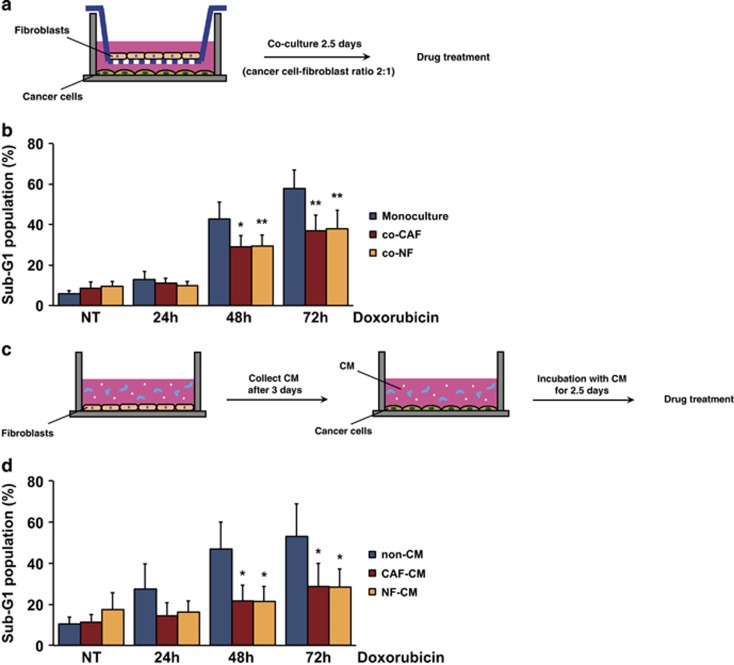
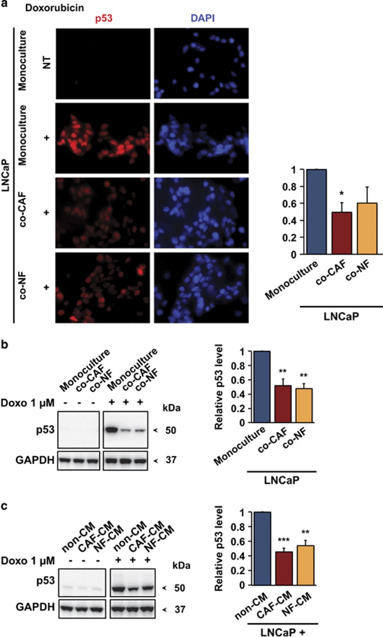
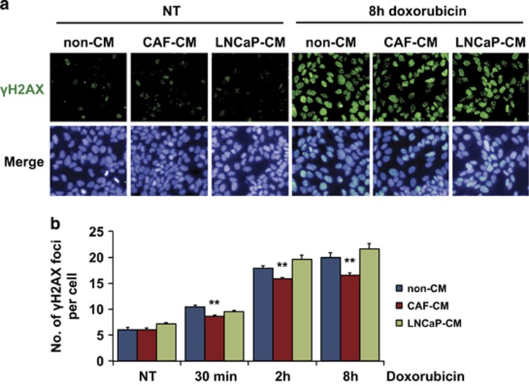
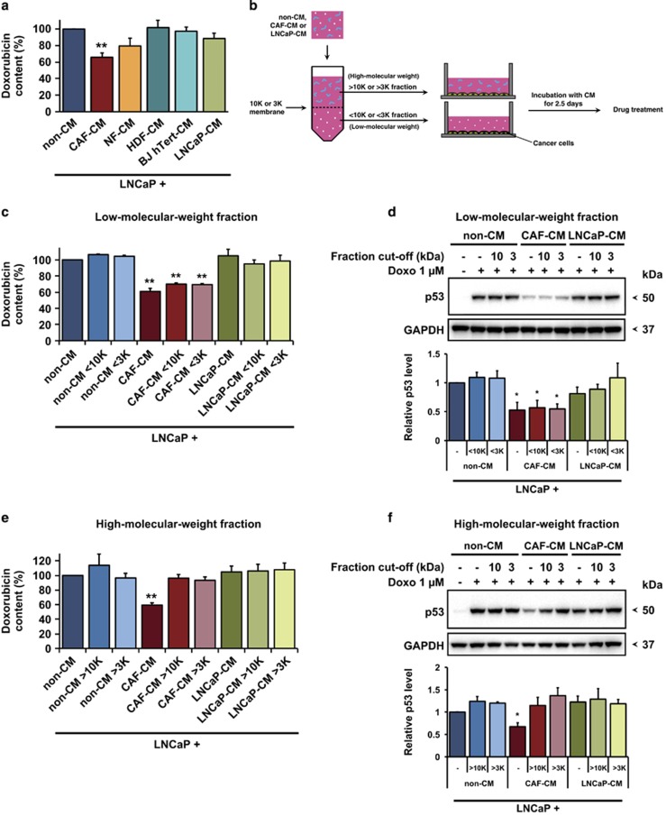
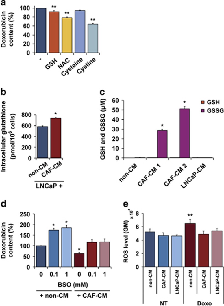
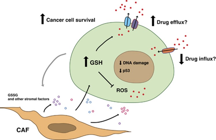
Drug resistance is a major problem in cancer therapy. A growing body of evidence demonstrates that the tumor microenvironment, including cancer-associated fibroblasts (CAFs), can modulate drug sensitivity in tumor cells. We examined the effect of primary human CAFs on p53 induction and cell viability in prostate cancer cells on treatment with chemotherapeutic drugs. Co-culture with prostate CAFs or CAF-conditioned medium attenuated DNA damage and the p53 response to chemotherapeutic drugs and enhanced prostate cancer cell survival. CAF-conditioned medium inhibited the accumulation of doxorubicin, but not taxol, in prostate cancer cells in a manner that was associated with increased cancer cell glutathione levels. A low molecular weight fraction (<3 kDa) of CAF-conditioned medium had the same effect. CAF-conditioned medium also inhibited induction of reactive oxygen species (ROS) in both doxorubicin- and taxol-treated cancer cells. Our findings suggest that CAFs can enhance drug resistance in cancer cells by inhibiting drug accumulation and counteracting drug-induced oxidative stress. This protective mechanism may represent a novel therapeutic target in cancer.


Conflict of interest statement

KGW and VJNB are co-founders and shareholders of Aprea Therapeutics AB, a company that develops novel p53-based cancer therapy. KGW is a member of its Clinical Advisory Board. The remaining authors declare no conflict of interest.

Figures

Figure 1
Figure 1
CAFs inhibit drug-induced LNCaP cell death. (a) Schematic of the experimental set-up for transwell co-culture assay. (b) FACS-PI analysis showing sub-G1 cell population of LNCaP cells co-cultured with fibroblasts (CAF or NF) after 24, 48 and 72 h of 1 μM doxorubicin treatment (mean and S.E.M.; N=7; *P<0.05, **P<0.01). (c) Schematic image of the conditioned medium (CM) assay. (d) Sub-G1 cell population of LNCaP cells grown with fresh (non-CM) or fibroblast- (CAF or NF) conditioned medium (CM), as determined by FACS-PI analysis after 24, 48 and 72 h exposure of 1 μM doxorubicin (mean and S.E.M.; N=5; *P<0.05). NT, non-treated
Figure 2
Figure 2
CAFs attenuate the induction of p53 in LNCaP cells. (a) Left, representative immunofluorescence staining of p53 (red) in LNCaP cells co-cultured with fibroblasts (CAF or NF) after 9 h of 1 μM doxorubicin treatment. Cell nuclei were stained with DAPI (blue). LNCaP-eGFP cells were used to distinguish LNCaP cells from the fibroblasts. Original magnification × 40. Right, quantitative graph of nuclear p53 staining after doxorubicin treatment in relation to p53 intensity in doxorubicin-treated monoculture (mean and S.E.M.; N=3; *P<0.05). NT, non-treated. (b) Left panel: p53 accumulation after 9 h of 1 μM doxorubicin treatment in LNCaP cells co-cultured with fibroblasts (CAF or NF) using transwell system, as indicated by a representative immunoblot of whole-cell lysates. Right panel: quantification of p53 level after drug treatment in relation to p53 level of doxorubicin-treated monoculture (mean and S.E.M.; N=6; **P<0.01). (c) Left panel: p53 accumulation in LNCaP cells after incubation with fresh (non-CM) or (CAF- or NF-) conditioned medium (CM) and 9 h of 1 μM doxorubicin treatment, as indicated by a representative immunoblot. Right panel: quantification of p53 level after the treatment in relation to p53 level of control treated with non-conditioned medium and doxorubicin (mean and S.E.M.; N=7; **P<0.01, ***P<0.0001)
Figure 3
Figure 3
Decreased DNA damage in LNCaP cells cultivated in CAF-CM. (a) Representative images of γH2AX foci (green) and nuclei (blue) visualized with Operetta, an automated fluorescence microscope, after 8 h of doxorubicin treatment. Original magnification × 20. (b) Average number of γH2AX foci per cell after 30 min, 2 h and 8 h of 1 μM doxorubicin treatment (mean and S.E.M.; N=3; **P<0.01). The foci numbers were obtained with Operetta and analyzed with Columbus software
Figure 4
Figure 4
The low molecular weight fraction of CAF-CM decreases doxorubicin accumulation in LNCaP cells. (a) Doxorubicin content assessed by FACS in LNCaP cells exposed to various conditioned media (CM) and 8 h of 1 μM doxorubicin (mean and S.E.M.; N=5; **P<0.01). (b) Schematic diagram of the separation of fresh (non-CM) or CAF- or LNCaP-conditioned medium (CM) into high- and low molecular weight fractions with cut-off of 10 kDa or 3 kDa. (c) Effect of low molecular weight fraction on doxorubicin accumulation, as assessed by FACS (mean and S.E.M.; N=3; **P<0.01) and (d) upper panel: p53 induction in LNCaP cells exposed to 8 h of 1 μM doxorubicin, as indicated by a representative immunoblot. Lower panel: quantification of p53 level after the treatment in relation to p53 level of control treated with non-conditioned medium and doxorubicin (mean and S.E.M.; N=4; *P<0.05). (e) Effect of high molecular weight fraction on intracellular doxorubicin level as assessed by FACS (mean and S.E.M.; N=3; **P<0.01) and (f) upper panel: p53 induction in LNCaP cells exposed to 1 μM doxorubicin for 8 h, as shown by a representative immunoblot. Lower panel: quantification of p53 level after the treatment in relation to p53 level of control treated with non-conditioned medium and doxorubicin (mean and S.E.M.; N=4; *P<0.05)
Figure 5
Figure 5
Role of glutathione and its precursors for doxorubicin accumulation in LNCaP cells. (a) Reduction in doxorubicin content in LNCaP cells after exposure to 5 mM reduced glutathione (GSH) or N-acetyl-l-cysteine (NAC) or 1 mM cysteine or cystine daily for 3 days, and 1 μM doxorubicin for 8 h, in relation to doxorubicin-treated only control (mean and S.E.M.; N=5; **P<0.01). (b) Total intracellular glutathione levels (GSH+GSSG) in LNCaP cells cultured in fresh (non-CM) or CAF-conditioned medium (CAF-CM), as determined by the glutathione assay described in the Materials and Methods' section (mean and S.E.M.; N=6; *P<0.05). (c) GSH and GSSG concentrations in different media assessed by HPLC (mean and S.E.M.; N=2; *P<0.05). Two different pools of CAF-conditioned medium were tested. (d) Effect of l-Buthionine Sulphoximine (BSO) on doxorubicin accumulation in LNCaP cells in the presence or absence of CAF-CM. BSO was added to the culture medium from day 1. After 3 days of culture with conditioned medium in the presence or absence of BSO as indicated, the cells were exposed to 1 μM doxorubicin for 8 h (mean and S.E.M.; N=3; *P<0.05). (e) ROS levels in LNCaP cells after 3 h treatment with 1 μM doxorubicin as assessed by CellROX and expressed as geometric mean fluorescence intensity (mean and S.E.M.; N=3; **P<0.01). The cells were cultured in fresh (non-CM), CAF-conditioned medium (CAF-CM) or LNCaP-conditioned medium (LNCaP-CM)
Figure 6
Figure 6
CAFs promote drug resistance and cancer cell survival. Model for interactions between CAFs and cancer cells that lead to enhanced drug resistance. CAFs provide tumor cells with oxidized glutathione as well as other stromal factors. The components are taken up by cancer cells, leading to elevated intracellular GSH levels, inhibition of drug accumulation and inhibition of reactive oxygen species (ROS). CAF-mediated reduction of drug accumulation could possibly be due to decreased influx and/or increased efflux. As a result, DNA damage and p53 induction are attenuated, and cancer cell survival is increased

References

    1. Hanahan D, Weinberg RA. The hallmarks of cancer. Cell 2000; 100: 57–70. - PubMed
    1. Hanahan D, Weinberg RA. Hallmarks of cancer: the next generation. Cell 2011; 144: 646–674. - PubMed
    1. Hanahan D, Coussens LM. Accessories to the crime: functions of cells recruited to the tumor microenvironment. Cancer Cell 2012; 21: 309–322. - PubMed
    1. Qiu W, Hu M, Sridhar A, Opeskin K, Fox S, Shipitsin M et al. No evidence of clonal somatic genetic alterations in cancer-associated fibroblasts from human breast and ovarian carcinomas. Nat Genet 2008; 40: 650–655. - PMC - PubMed
    1. Allinen M, Beroukhim R, Cai L, Brennan C, Lahti-Domenici J, Huang H et al. Molecular characterization of the tumor microenvironment in breast cancer. Cancer Cell 2004; 6: 17–32. - PubMed

Publication types

MeSH terms