. 2017 Aug;44(8):4252-4261.
doi: 10.1002/mp.12391. Epub 2017 Jul 10.

A clinically validated prediction method for facial soft-tissue changes following double-jaw surgery

Affiliations

A clinically validated prediction method for facial soft-tissue changes following double-jaw surgery

Daeseung Kim et al. Med Phys. 2017 Aug.

Abstract

Purpose: It is clinically important to accurately predict facial soft-tissue changes prior to orthognathic surgery. However, the current simulation methods are problematic, especially in anatomic regions of clinical significance, e.g., the nose, lips, and chin. We developed a new 3-stage finite element method (FEM) approach that incorporates realistic tissue sliding to improve such prediction.

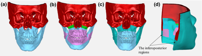
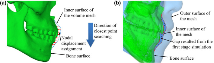
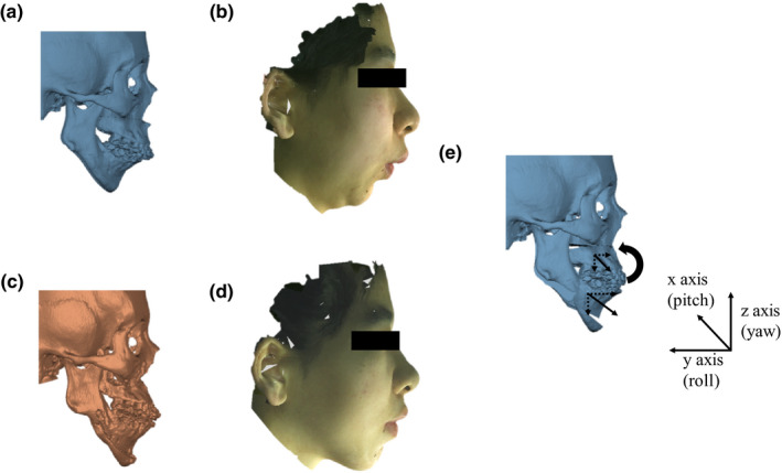
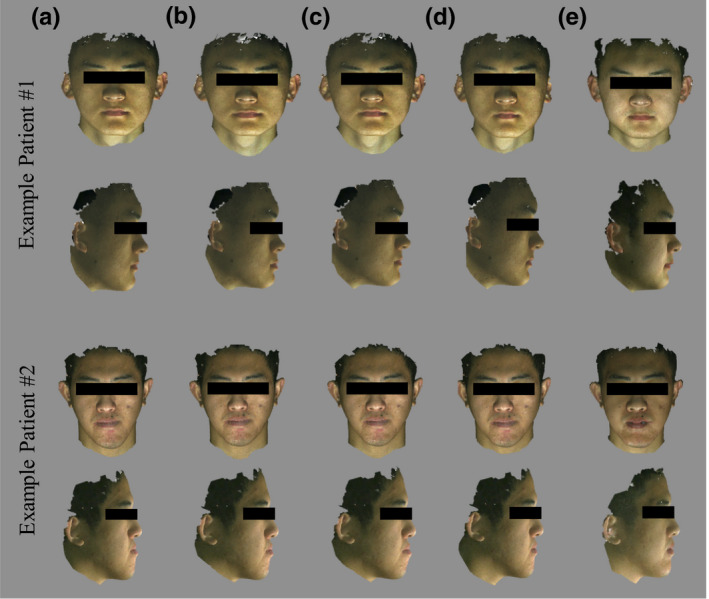
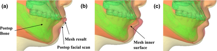
Methods: In Stage One, soft-tissue change was simulated, using FEM with patient-specific mesh models generated from our previously developed eFace template. Postoperative bone movement was applied on the patient mesh model with standard FEM boundary conditions. In Stage Two, the simulation was improved by implementing sliding effects between gum tissue and teeth using a nodal force constraint scheme. In Stage Three, the result of the tissue sliding effect was further enhanced by reassigning the soft-tissue-bone mapping and boundary conditions using nodal spatial constraint. Finally, our methods have been quantitatively and qualitatively validated using 40 retrospectively evaluated patient cases by comparing it to the traditional FEM method and the FEM with sliding effect, using a nodal force constraint method.

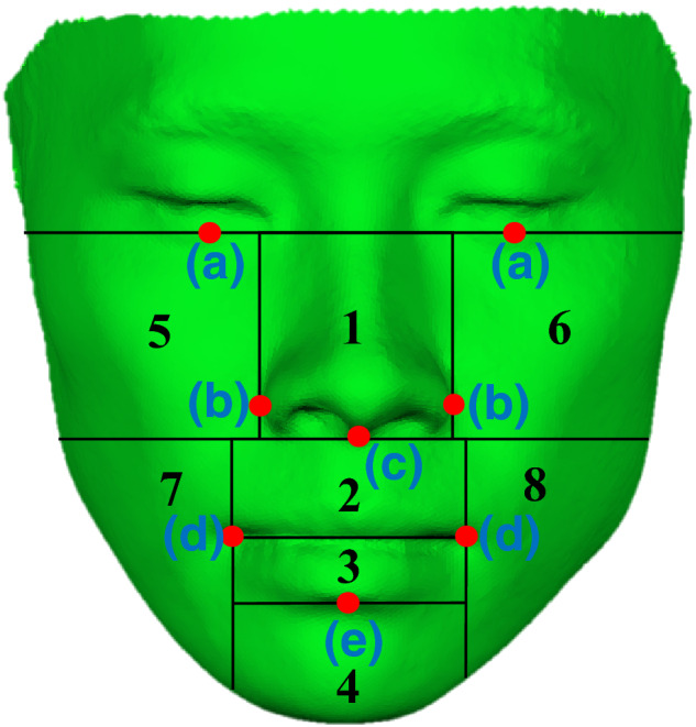
Results: The results showed that our method was better than the other two methods. Using our method, the quantitative distance errors between predicted and actual patient surfaces for the entire face and any subregions thereof were below 1.5 mm. The overall soft-tissue change prediction was accurate to within 1.1 ± 0.3 mm, with the accuracy around the upper and lower lip regions of 1.2 ± 0.7 mm and 1.5 ± 0.7 mm, respectively. The results of qualitative evaluation completed by clinical experts showed an improvement of 46% in acceptance rate compared to the traditional FEM simulation. More than 80% of the result of our approach was considered acceptable in comparison with 55% and 50% following the other two methods.

Conclusion: The FEM simulation method with improved sliding effect showed significant accuracy improvement in the whole face and the clinically significant regions (i.e., nose and lips) in comparison with the other published FEM methods, with or without sliding effect using a nodal force constraint. The qualitative validation also proved the clinical feasibility of the developed approach.

Keywords: facial soft-tissue change prediction; finite element model; orthognathic surgery; soft-tissue modeling; soft-tissue sliding effect.


Conflict of interest statement

The authors have no COI to report.

Figures

Figure 1
Figure 1
Mesh nodal boundary condition. (a) Mesh inner surface boundary condition (illustrated on the skull) for the first stage only; (b) Mesh inner surface boundary condition for the second stage only; (c) Mesh inner surface boundary condition (illustrated on the skull) for the third stage only. (d) Posterior and superior surface boundary condition for all stages. (Note that the inferoposterior regions are the fixed nodes in the first and third stage); Color coding of fixed nodes: red (or dimgray in B&W); Moving nodes: Blue (or lightgray in B&W); Sliding nodes: pink (or silver in B&W); Free nodes: GreenBlue (or gray in B&W); Tenting of periosteum: green (or gray in B&W). [Color figure can be viewed at wileyonlinelibrary.com]
Figure 2
Figure 2
Assigning nodal spatial constraint. (a) Description of nodal displacement boundary condition assignment in the third stage. (b) Mismatch between the simulated mesh inner surface and the bone surface after sliding effect using nodal force constraint. [Color figure can be viewed at wileyonlinelibrary.com]
Figure 3
Figure 3
Computing bony segment movements for soft‐tissue change prediction. (a) Preoperative positions of maxillary, mandibular, and chin segments. (b) Corresponding preoperative facial 3D color photograph of the preoperative facial soft tissue. (c) Postoperative position of the maxillary, mandibular, and chin segments. (d) Corresponding postoperative facial 3D color photograph of the preoperative facial soft tissue. (e) Computed surgical movements of the bony segments by registering preoperative bony segments to postoperative ones. Computed movement vectors of bony segments by first registering the postoperative CT models to the preoperative CT models at surgically unchanged regions (i.e., cranium and midface), then registering each virtually osteotomized preoperative bony segment from its original position to the postoperative one. Arrows represent the movements of the bony segment relative to the surgically unaltered cranium and midface. The coordinate system follows the right‐hand rule. [Color figure can be viewed at wileyonlinelibrary.com]
Figure 4
Figure 4
Sub‐regions (automatically divided using anatomical landmarks). The anatomical landmarks were: (a) palpebrale inferius, (b) alar, (c) subnasale, (d) cheilion, and (e) labrale inferius. [Color figure can be viewed at wileyonlinelibrary.com]
Figure 5
Figure 5
Randomly selected two examples of simulated results with color texture mapped. The top row shows the frontal view while the bottom row shows the right view for each example patient. (a) Preoperative soft tissue. (b) Predicted soft‐tissue change using Method #1. (c) Predicted soft‐tissue change using Method #2. (d) Predicted soft‐tissue change using Method #3, our complete 3‐stage approach. (e) The actual postoperative soft tissue. For the Example Patient #1, the quantitative analysis showed 7.8 and 6.5 mm of maximum displacement error for Methods #1 and #2 in the chin region, respectively, while that of Method #3 was 1.0 mm. However, the upper and lower lip relationship resulted from the Method #3 was only getting similar to the actual postoperative lip relationship, even though the quantitative error was significantly improved. For the Example Patient #2, the quantitative analysis showed 3.6 and 5.3 mm of maximum displacement error for Methods #1 and #2 in the lower lip region, respectively, while that of Method #3 was 3.2 mm. The result achieved with Method #3 is the only one showing correct upper and lower lip relationship comparing to the actual postoperative soft tissue, even though the quantitative improvement was not significant. [Color figure can be viewed at wileyonlinelibrary.com]
Figure 6
Figure 6
An example of quantitative and qualitative evaluation results. The predicted mesh (red (or gray in B&W)) is superimposed onto the postoperative bone (green (or dimgray in B&W)) and soft tissue (light yellow (or lightgray in B&W)). (a) The predicted result using Method #1. (b) The predicted result using Method #2. (c) The predicted result using Method #3. [Color figure can be viewed at wileyonlinelibrary.com]

References

    1. Xia JJ, Gateno J, Teichgraeber JF, et al. Algorithm for planning a double‐jaw orthognathic surgery using a computer‐aided surgical simulation (CASS) protocol. Part 1: planning sequence. Int J Oral Maxillofac Surg. 2015;44:1431–1440. - PMC - PubMed
    1. McCormick SU, Drew SJ. Virtual model surgery for efficient planning and surgical performance. J Oral Maxillofac Surg. 2011;69:638–644. - PubMed
    1. Bobek S, Farrell B, Choi C, Farrell B, Weimer K, Tucker M. Virtual surgical planning for orthognathic surgery using digital data transfer and an intraoral fiducial marker: the charlotte method. J Oral Maxillofac Surg. 2015;73:1143–1158. - PubMed
    1. Bell WH. Modern Practice in Orthognathic and Reconstructive Surgery: volume 3. Philadelphia, PA: W.B. Saunders company; 1993.
    1. Chabanas M, Luboz V, Payan Y. Patient specific finite element model of the face soft tissues for computer‐assisted maxillofacial surgery. Med Image Anal. 2003;7:131–151. - PubMed

LinkOut - more resources