

Classification of breast cancer histology images using Convolutional Neural Networks
- PMID: 28570557
- PMCID: PMC5453426
- DOI: 10.1371/journal.pone.0177544
Classification of breast cancer histology images using Convolutional Neural Networks
Abstract
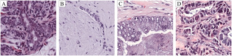
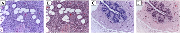
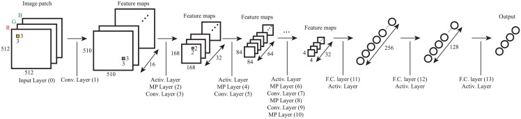
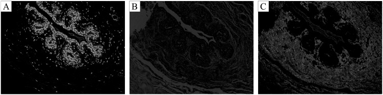
Breast cancer is one of the main causes of cancer death worldwide. The diagnosis of biopsy tissue with hematoxylin and eosin stained images is non-trivial and specialists often disagree on the final diagnosis. Computer-aided Diagnosis systems contribute to reduce the cost and increase the efficiency of this process. Conventional classification approaches rely on feature extraction methods designed for a specific problem based on field-knowledge. To overcome the many difficulties of the feature-based approaches, deep learning methods are becoming important alternatives. A method for the classification of hematoxylin and eosin stained breast biopsy images using Convolutional Neural Networks (CNNs) is proposed. Images are classified in four classes, normal tissue, benign lesion, in situ carcinoma and invasive carcinoma, and in two classes, carcinoma and non-carcinoma. The architecture of the network is designed to retrieve information at different scales, including both nuclei and overall tissue organization. This design allows the extension of the proposed system to whole-slide histology images. The features extracted by the CNN are also used for training a Support Vector Machine classifier. Accuracies of 77.8% for four class and 83.3% for carcinoma/non-carcinoma are achieved. The sensitivity of our method for cancer cases is 95.6%.
Conflict of interest statement
Figures
Similar articles
-
Computer assisted recognition of breast cancer in biopsy images via fusion of nucleus-guided deep convolutional features.Comput Methods Programs Biomed. 2020 Oct;194:105531. doi: 10.1016/j.cmpb.2020.105531. Epub 2020 May 11. Comput Methods Programs Biomed. 2020. PMID: 32422473
-
Deep Convolutional Neural Networks for breast cancer screening.Comput Methods Programs Biomed. 2018 Apr;157:19-30. doi: 10.1016/j.cmpb.2018.01.011. Epub 2018 Jan 11. Comput Methods Programs Biomed. 2018. PMID: 29477427
-
Detection of Breast Cancer with Lightweight Deep Neural Networks for Histology Image Classification.Crit Rev Biomed Eng. 2022;50(2):1-19. doi: 10.1615/CritRevBiomedEng.2022043417. Crit Rev Biomed Eng. 2022. PMID: 36374820
-
Involvement of Machine Learning for Breast Cancer Image Classification: A Survey.Comput Math Methods Med. 2017;2017:3781951. doi: 10.1155/2017/3781951. Epub 2017 Dec 31. Comput Math Methods Med. 2017. PMID: 29463985 Free PMC article. Review.
-
Breast cancer cell nuclei classification in histopathology images using deep neural networks.Int J Comput Assist Radiol Surg. 2018 Feb;13(2):179-191. doi: 10.1007/s11548-017-1663-9. Epub 2017 Aug 31. Int J Comput Assist Radiol Surg. 2018. PMID: 28861708 Review.
Cited by
-
Development and evaluation of a deep neural network for histologic classification of renal cell carcinoma on biopsy and surgical resection slides.Sci Rep. 2021 Mar 29;11(1):7080. doi: 10.1038/s41598-021-86540-4. Sci Rep. 2021. PMID: 33782535 Free PMC article.
-
A multi-phase deep CNN based mitosis detection framework for breast cancer histopathological images.Sci Rep. 2021 Mar 18;11(1):6215. doi: 10.1038/s41598-021-85652-1. Sci Rep. 2021. PMID: 33737632 Free PMC article.
-
Impact of Imaging Biomarkers and AI on Breast Cancer Management: A Brief Review.Cancers (Basel). 2023 Oct 30;15(21):5216. doi: 10.3390/cancers15215216. Cancers (Basel). 2023. PMID: 37958390 Free PMC article. Review.
-
Deep Learning Classification of Breast Cancer Tissue from Terahertz Imaging Through Wavelet Synchro-Squeezed Transformation and Transfer Learning.J Infrared Millim Terahertz Waves. 2022 Jan;43(1-2):48-70. doi: 10.1007/s10762-021-00839-x. J Infrared Millim Terahertz Waves. 2022. PMID: 36246840 Free PMC article.
-
Classification of Multiple H&E Images via an Ensemble Computational Scheme.Entropy (Basel). 2023 Dec 28;26(1):34. doi: 10.3390/e26010034. Entropy (Basel). 2023. PMID: 38248160 Free PMC article.
References
-
- Siegel RL, Miller KD, Jemal A. Cancer statistics, 2016. CA: A Cancer Journal for Clinicians. 2016;66(1):7–30. - PubMed
-
- Smith Ra, Cokkinides V, Eyre HJ. American Cancer Society Guidelines for the Early Detection of Cancer, 2004. CA: A Cancer Journal for Clinicians. 2004;54(1):41–52. - PubMed
-
- Pêgo A, Aguiar P. Bioimaging 2015; 2015. Available from: http://www.bioimaging2015.ineb.up.pt/dataset.html.
-
- NationalBreastCancerFoundation. Breast Cancer Diagnosis; 2015. Available from: http://www.nationalbreastcancer.org/breast-cancer-diagnosis.
MeSH terms
LinkOut - more resources
Full Text Sources
Other Literature Sources
Medical
