. 2017 Jun 1;12(6):e0178193.
doi: 10.1371/journal.pone.0178193. eCollection 2017.

Characteristics of HIV target CD4 T cells collected using different sampling methods from the genital tract of HIV seronegative women

Affiliations

Characteristics of HIV target CD4 T cells collected using different sampling methods from the genital tract of HIV seronegative women

Smita S Iyer et al. PLoS One. .

Abstract

Background: Understanding the immune profile of CD4 T cells, the primary targets for HIV, in the female genital tract (FGT) is critical for evaluating and developing effective biomedical HIV prevention strategies in women. However, longitudinal investigation of HIV susceptibility markers expressed by FGT CD4 T cells has been hindered by low cellular yield and risk of sampling-associated trauma. We investigated three minimally invasive FGT sampling methods to characterize and compare CD4 T cell yield and phenotype with the goal of establishing feasible sampling strategies for immune profiling of mucosal CD4 T cells.

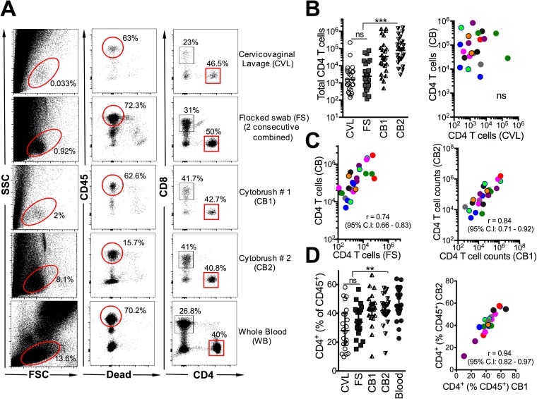
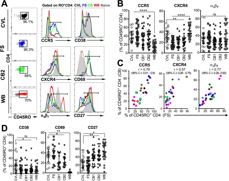
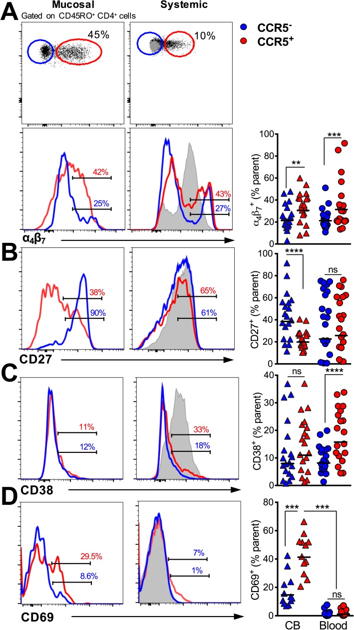
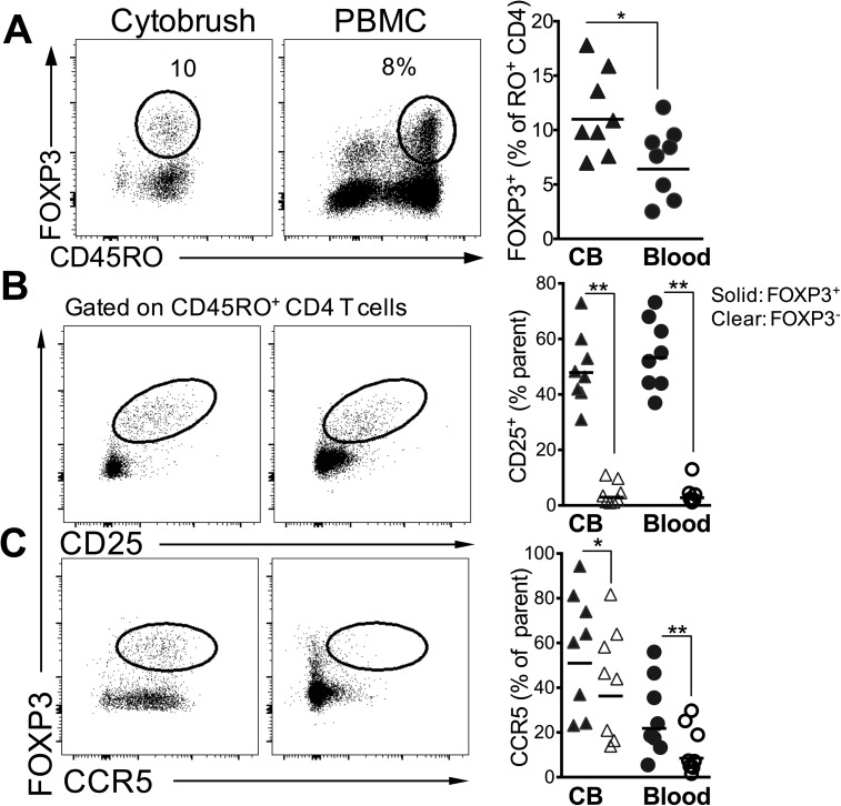
Methods and results: FGT samples were collected bimonthly from 12 healthy HIV negative women of reproductive age in the following order: 1) Cervicovaginal lavage (CVL), 2) two sequential endocervical flocked swabs (FS), and 3) two sequential endocervical cytobrushes (CB1, CB2). Cells were isolated and phentoyped via flow cytometry. CD4 T cell recovery was highest from each individual CB compared to either CVL or FS (p < 0.0001). The majority of CD4 T cells within the FGT, regardless of sampling method, expressed CCR5 relative to peripheral blood (p < 0.01). Within the CB, CCR5+ CD4 T cells expressed significantly higher levels of α4β7, CD69, and low levels of CD27 relative to CCR5- CD4 T cells (all p < 0.001). We also identified CD4 Treg lineage cells expressing CCR5 among CB samples.

Conclusions: Using three different mucosal sampling methods collected longitudinally we demonstrate that CD4 T cells within the FGT express CCR5 and α4β7 and are highly activated, attributes which could act in concert to facilitate HIV acquisition. FS and CB sampling methods can allow for investigation of strategies to reduce HIV target cells in the FGT and could inform the design and interpretation microbicide and vaccine studies in women.


Conflict of interest statement

Competing Interests: The authors have declared that no competing interests exist.

Figures

Fig 1
Fig 1. Highest CD4 T cell recovery from endocervical cytobrushes relative to endocervical flocked swabs and cervicovaginal lavages.
(A) Example analysis of human genital mucosal specimens from 9 women sequentially sampled for cervicovaginal lavage (CVL), flocked swab (FS) followed by two consecutive cytobrushes (CB1, CB2). The cellular components were isolated by centrifugation and incubated with antibodies to identify T lymphocyte subsets at 4°C for 30 minutes. Whole blood from the same individual was stained as a positive control. Scatter plots show data for nine participants over two-three independent visits at either the follicular or the luteal phase of the menstrual cycle. Participants are color coded for correlation plots. (B) CD4 T cell yields significantly higher from CB sampling relative to either CVL or FS, CD4 T cell yields from CB are not associated with yields from CVL. (C) CD4 T cell yields from FS correlate with CB yields, and CB1 and CB2 yields strongly correlate with each other. (D) CD4 T cell distribution profile across sampling methods, frequency of CD4+ cells similar and correlated between CB1 and CB2. ***, p < 0.001; **, p < 0.01, *, p < 0.05.
Fig 2
Fig 2. Enrichment of CD4 T cells expressing CCR5 and markers of activation in genital mucosa relative to blood.
(A) Representative flow plots show that CD4 T cells from CVL, FS and CB are highly enriched for antigen-experienced CD4 T cells as evidenced by expression of CD45RO. Histograms show comparison of expression of CCR5, CD38, CXCR4, CD69, α4β7 and CD27 on genital and whole blood (WB) CD4 T cells. Naive CD4 T cells are overlaid in grey. (B) Distribution of HIV co-receptors CCR5 (n = 16 visits for CVL, 24 visits for FS, CB1, CB2, and WB), CXCR4 (n = 19 visits for CVL, 21 visits for FS, 22 visits for CB1, CB2, and WB), and expression of the integrin α4β7 (n = 22 visits for CVL, 27 visits for FS, CB1, CB2, and WB) on mucosal and blood CD4 T cells. (C) Correlation of CCR5, CXCR4 and α4β7 expression on FS and CB. (D) Distribution of activation markers CD38 (n = 21 visits for CVL, 25 visits for FS, CB1, CB2, and WB), CD69 (n = 14 visits for CVL, 15 visits for FS, CB1, CB2, and WB) and CD27 (n = 19 visits for CVL, 23 visits for FS, CB1, CB2, and WB) on mucosal and blood CD4 T cells (****, p < 0.0001; **, p < 0.01; *, p < 0.05).
Fig 3
Fig 3. HIV target CCR5+ CD4 T cells in female genital mucosa display phenotypic attributes of activation and trafficking to rectal mucosa.
Histograms show comparison of expression of markers between CD45RO+ CCR5+ and CCR5- cells genital and whole blood CD4 T cells for (A) α4β7, (B) CD27, (C) CD38 and (D) CD69 (***, p < 0.001, n = 20 participant visits for all markers except CD69 for which n = 12 visits)
Fig 4
Fig 4. Frequency and phenotype of CD4 T Regulatory cells in the genital mucosa.
(A) Representative flow plots showing FOXP3+ CD4 T cells in cytobrush and PBMCs (plots are gated on total CD4+ T cells). Scatter plot shows frequency of Tregs in 8 participants. (B) shows expression of CD25 and (C) shows CCR5 expression in FOXP3+ and FOXP3- CD45RO+ cells (**, p < 0.01; *, p < 0.05 using a two-tailed, paired non-parametric t test

References

    1. Mestecky J, Moldoveanu Z, Smith PD, Hel Z, Alexander RC. Mucosal immunology of the genital and gastrointestinal tracts and HIV-1 infection. Journal of reproductive immunology. 2009;83(1–2):196–200. Epub 2009/10/27. PubMed Central PMCID: PMC2802574. doi: 10.1016/j.jri.2009.07.005 - DOI - PMC - PubMed
    1. Chen NE, Meyer JP, Springer SA. Advances in the prevention of heterosexual transmission of HIV/AIDS among women in the United States. Infectious disease reports. 2011;3(1). Epub 2011/01/01. PubMed Central PMCID: PMC3671603. - PMC - PubMed
    1. McKinnon LR, Kaul R. Quality and quantity: mucosal CD4+ T cells and HIV susceptibility. Current opinion in HIV and AIDS. 2012;7(2):195–202. Epub 2012/02/09. doi: 10.1097/COH.0b013e3283504941 - DOI - PubMed
    1. Hladik F, Sakchalathorn P, Ballweber L, Lentz G, Fialkow M, Eschenbach D, et al. Initial events in establishing vaginal entry and infection by human immunodeficiency virus type-1. Immunity. 2007;26(2):257–70. PubMed Central PMCID: PMCPMC1885958. doi: 10.1016/j.immuni.2007.01.007 - DOI - PMC - PubMed
    1. Yi TJ, Shannon B, Prodger J, McKinnon L, Kaul R. Genital immunology and HIV susceptibility in young women. Am J Reprod Immunol. 2013;69 Suppl 1:74–9. Epub 2012/11/20. - PubMed