. 2017 Jun 1;12(6):e0178700.
doi: 10.1371/journal.pone.0178700. eCollection 2017.

Frameshift indels introduced by genome editing can lead to in-frame exon skipping

Affiliations

Frameshift indels introduced by genome editing can lead to in-frame exon skipping

Simon Lalonde et al. PLoS One. .

Abstract

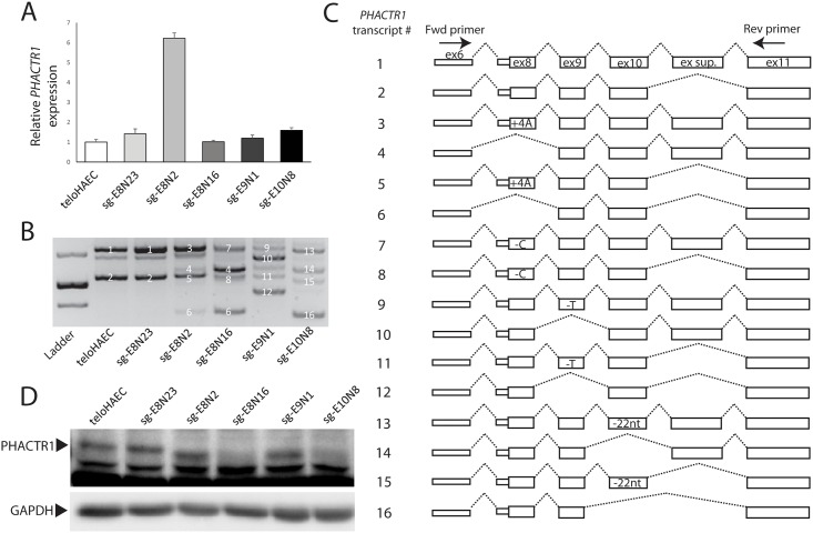
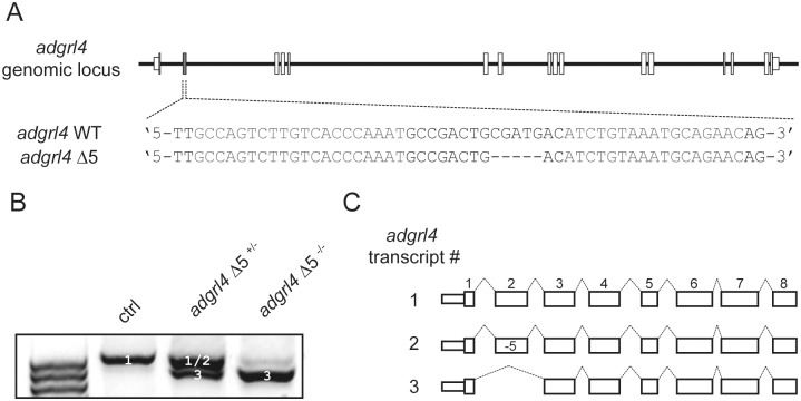
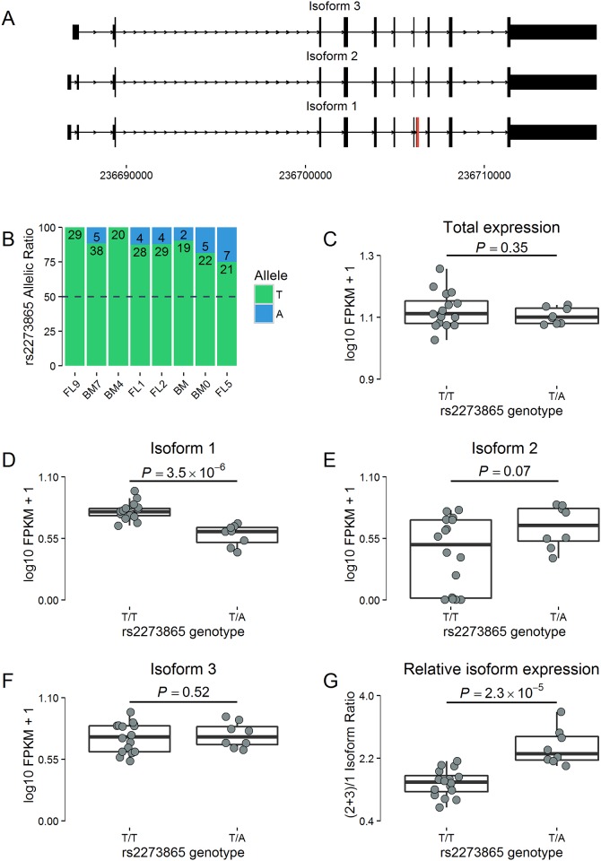
The introduction of frameshift indels by genome editing has emerged as a powerful technique to study the functions of uncharacterized genes in cell lines and model organisms. Such mutations should lead to mRNA degradation owing to nonsense-mediated mRNA decay or the production of severely truncated proteins. Here, we show that frameshift indels engineered by genome editing can also lead to skipping of "multiple of three nucleotides" exons. Such splicing events result in in-frame mRNA that may encode fully or partially functional proteins. We also characterize a segregating nonsense variant (rs2273865) located in a "multiple of three nucleotides" exon of LGALS8 that increases exon skipping in human erythroblast samples. Our results highlight the potentially frequent contribution of exonic splicing regulatory elements and are important for the interpretation of negative results in genome editing experiments. Moreover, they may contribute to a better annotation of loss-of-function mutations in the human genome.


Conflict of interest statement

Competing Interests: The authors have declared that no competing interests exist.

Figures

Fig 1
Fig 1. Frameshift indels cause in-frame exon skipping in PHACTR1.
(A) PHACTR1 expression levels measured by real-time qPCR in the parental teloHAEC cell line, an unedited clone (sg-E8N23), and clones with CRISPR-Cas9-generated frameshift indels in PHACTR1 exon 8 (sg-E8N2 and sg-E8N16), exon 9 (sg-E9N1), and exon 10 (sg-E10N8). Data show mean and standard error of the mean from two biological replicates, done in triplicates. PHACTR1 expression levels in sg-E8N2 is 6.2 fold greater than in the parental teloHAEC cell line (Student’s t-test P = 0.0033). (B) Agarose gel electrophoresis profile of the main PHACTR1 isoforms detected in cDNA from teloHAEC cells, unedited clones, or clones with a frameshift indel. We assigned a transcript number to each of the PHACTR1 isoform that we could Sanger sequence and align to the reference sequence. Unlabeled bands could not be assigned to PHACTR1. Bands in the molecular ladder correspond to 400, 500 and 700-bp. This gel is representative of three independent experiments. (C) Schematic diagram of all the PHACTR1 isoforms that we identified in the different teloHAEC cell lines. Transcript numbers correspond to the bands (white numbers) in B. The PCR primers in exon 6 and 11 are depicted. For the isoforms expressed in edited clones, we added the corresponding nucleotide changes introduced by the frameshift indels. (D) Western blot of PHACTR1 in the parental, unedited and edited teloHAEC cells. The arrowhead indicates PHACTR1, lower bands are non-specific proteins recognized by the antibody. PHACTR1 is smaller in sg-E8N2, consistent with skipping of exon 8 or usage of an alternative in-frame start codon downstream of the frameshift indel. For sg-E9N1, the smaller PHACTR1 protein is consistent with skipping of exon 9. We could not detect PHACTR1 proteins in sg-E8N16 and sg-E10N8. We used GAPDH as loading control. This Western blot is representative of three independent experiments.
Fig 2
Fig 2. Frameshift indel can lead to exon skipping in the zebrafish adgrl4 gene.
(A) adgrl4 genomic locus, TALEN target site in exon 2 of the adgrl4 gene and a stable mutant line (Δ5) that was analyzed. (B) PCR analysis of adgrl4 mRNA transcripts in 10 to 15 pooled embryo samples from control (ctrl), adgrl4 Δ5+/-, and adgrl4 Δ5-/- fishes. (C) Schematic representation of the different transcripts recovered from the bands (white numbers) in B.
Fig 3
Fig 3. Exon skipping in LGALS8 in human erythroblasts.
(A) In in vitro differentiated human erythroblasts, three LGALS8 mRNA isoforms are expressed. Isoform 1 includes the “multiple of three nucleotides” exon 9 (in red, 126-bp), whereas isoforms 2 and 3 do not. The nonsense variant rs2273865 (p.Leu212Ter) is located in exon 9. At this variant, the minor A-allele has a frequency of 3.5% in populations of European ancestry (ExAC). (B) Eight erythroblast samples are heterozygous at rs2273865 and show strong allelic imbalance (binomial P<0.05 for all samples). Numbers in the bars indicate the numbers of reads carrying the T (green) or A (blue) allele. Differential expression of total LGALS8 (C), isoform 1 (D), isoform 2 (E), and isoform 3 (F) between erythroblast samples homozygous TT (n = 16) and heterozygous AT (n = 8) at rs2273865. No samples homozygous for the minor allele (AA) were available. (G) The ratio of LGALS8 transcripts without exon 9 over transcripts with exon 9 is higher in heterozygous AT than in homozygous TT erythroblast samples. P-values are calculated by linear regression correcting for cell developmental stage.

References

    1. Gupta RM, Musunuru K. Expanding the genetic editing tool kit: ZFNs, TALENs, and CRISPR-Cas9. The Journal of clinical investigation. 2014;124(10):4154–61. 10.1172/JCI72992 ; - DOI - PMC - PubMed
    1. Wright AV, Nunez JK, Doudna JA. Biology and Applications of CRISPR Systems: Harnessing Nature's Toolbox for Genome Engineering. Cell. 2016;164(1–2):29–44. 10.1016/j.cell.2015.12.035 . - DOI - PubMed
    1. Maquat LE. When cells stop making sense: effects of nonsense codons on RNA metabolism in vertebrate cells. RNA. 1995;1(5):453–65. ; - PMC - PubMed
    1. Wang T, Wei JJ, Sabatini DM, Lander ES. Genetic screens in human cells using the CRISPR-Cas9 system. Science. 2014;343(6166):80–4. 10.1126/science.1246981 ; - DOI - PMC - PubMed
    1. Shalem O, Sanjana NE, Hartenian E, Shi X, Scott DA, Mikkelsen TS, et al. Genome-scale CRISPR-Cas9 knockout screening in human cells. Science. 2014;343(6166):84–7. 10.1126/science.1247005 ; - DOI - PMC - PubMed