

Thin silica shell coated Ag assembled nanostructures for expanding generality of SERS analytes
- PMID: 28570633
- PMCID: PMC5453564
- DOI: 10.1371/journal.pone.0178651
Thin silica shell coated Ag assembled nanostructures for expanding generality of SERS analytes
Abstract
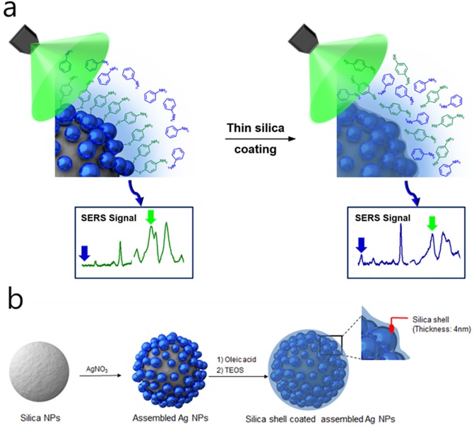
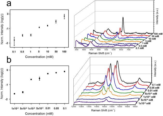
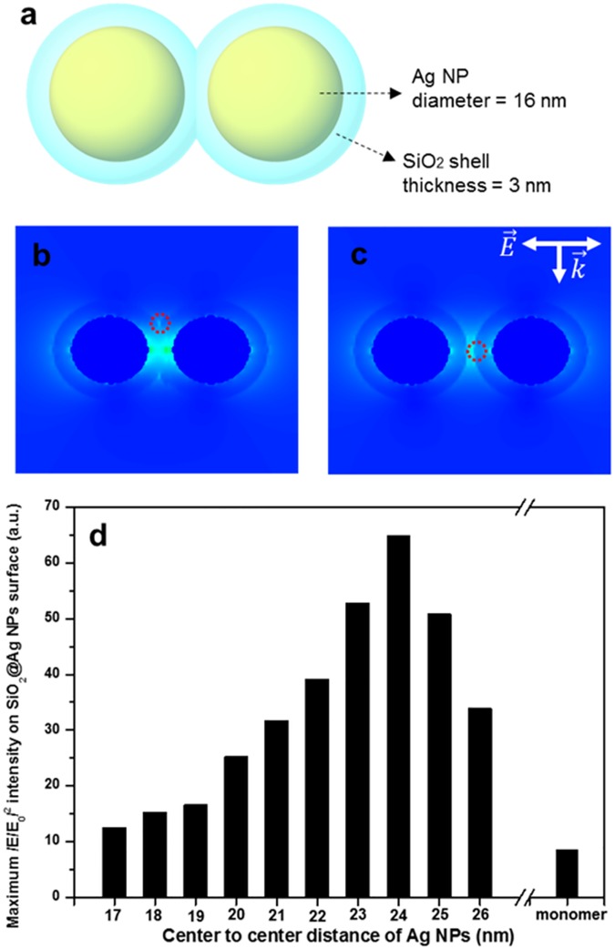
Surface-enhanced Raman scattering (SERS) provides a unique non-destructive spectroscopic fingerprint for chemical detection. However, intrinsic differences in affinity of analyte molecules to metal surface hinder SERS as a universal quantitative detection tool for various analyte molecules simultaneously. This must be overcome while keeping close proximity of analyte molecules to the metal surface. Moreover, assembled metal nanoparticles (NPs) structures might be beneficial for sensitive and reliable detection of chemicals than single NP structures. For this purpose, here we introduce thin silica-coated and assembled Ag NPs (SiO2@Ag@SiO2 NPs) for simultaneous and quantitative detection of chemicals that have different intrinsic affinities to silver metal. These SiO2@Ag@SiO2 NPs could detect each SERS peak of aniline or 4-aminothiophenol (4-ATP) from the mixture with limits of detection (LOD) of 93 ppm and 54 ppb, respectively. E-field distribution based on interparticle distance was simulated using discrete dipole approximation (DDA) calculation to gain insight into enhanced scattering of these thin silica coated Ag NP assemblies. These NPs were successfully applied to detect aniline in river water and tap water. Results suggest that SiO2@Ag@SiO2 NP-based SERS detection systems can be used as a simple and universal detection tool for environment pollutants and food safety.
Conflict of interest statement
Figures

Similar articles
-
Control of Silver Coating on Raman Label Incorporated Gold Nanoparticles Assembled Silica Nanoparticles.Int J Mol Sci. 2019 Mar 13;20(6):1258. doi: 10.3390/ijms20061258. Int J Mol Sci. 2019. PMID: 30871136 Free PMC article.
-
Ultra-Fine Control of Silica Shell Thickness on Silver Nanoparticle-Assembled Structures.Int J Mol Sci. 2021 Nov 5;22(21):11983. doi: 10.3390/ijms222111983. Int J Mol Sci. 2021. PMID: 34769413 Free PMC article.
-
"Elastic" property of mesoporous silica shell: for dynamic surface enhanced Raman scattering ability monitoring of growing noble metal nanostructures via a simplified spatially confined growth method.ACS Appl Mater Interfaces. 2015 Apr 15;7(14):7516-25. doi: 10.1021/acsami.5b01077. Epub 2015 Apr 3. ACS Appl Mater Interfaces. 2015. PMID: 25815901
-
Synthesis of MBA-Encoded Silver/Silica Core-Shell Nanoparticles as Novel SERS Tags for Biosensing Gibberellin A3 Based on Au@Fe3O4 as Substrate.Sensors (Basel). 2019 Nov 25;19(23):5152. doi: 10.3390/s19235152. Sensors (Basel). 2019. PMID: 31775290 Free PMC article.
-
Growth of Spherical Gold Satellites on the Surface of Au@Ag@SiO2 Core-Shell Nanostructures Used for an Ultrasensitive SERS Immunoassay of Alpha-Fetoprotein.ACS Appl Mater Interfaces. 2019 Jan 23;11(3):3617-3626. doi: 10.1021/acsami.8b21238. Epub 2019 Jan 11. ACS Appl Mater Interfaces. 2019. PMID: 30608142
Cited by
-
Prospects of Surface-Enhanced Raman Spectroscopy for Biomarker Monitoring toward Precision Medicine.ACS Photonics. 2022 Feb 16;9(2):333-350. doi: 10.1021/acsphotonics.1c01934. Epub 2022 Feb 2. ACS Photonics. 2022. PMID: 35211644 Free PMC article. Review.
-
Surface-Enhanced Raman Spectroscopy on Self-Assembled Au Nanoparticles Arrays for Pesticides Residues Multiplex Detection under Complex Environment.Nanomaterials (Basel). 2019 Mar 13;9(3):426. doi: 10.3390/nano9030426. Nanomaterials (Basel). 2019. PMID: 30871181 Free PMC article.
-
Functionalized β-Cyclodextrin Immobilized on Ag-Embedded Silica Nanoparticles as a Drug Carrier.Int J Mol Sci. 2019 Jan 14;20(2):315. doi: 10.3390/ijms20020315. Int J Mol Sci. 2019. PMID: 30646562 Free PMC article.
-
Silver Nano/Microparticles: Modification and Applications.Int J Mol Sci. 2019 May 28;20(11):2609. doi: 10.3390/ijms20112609. Int J Mol Sci. 2019. PMID: 31141905 Free PMC article.
-
Mono-6-Deoxy-6-Aminopropylamino-β-Cyclodextrin on Ag-Embedded SiO2 Nanoparticle as a Selectively Capturing Ligand to Flavonoids.Nanomaterials (Basel). 2019 Sep 20;9(10):1349. doi: 10.3390/nano9101349. Nanomaterials (Basel). 2019. PMID: 31547075 Free PMC article.
References
-
- Hering K, Cialla D, Ackermann K, Doerfer T, Moeller R, Schneidewind H, et al. SERS: a versatile tool in chemical and biochemical diagnostics. Analytical and Bioanalytical Chemistry. 2008;390(1):113–24. doi: 10.1007/s00216-007-1667-3 - DOI - PubMed
-
- Kudelski A. Analytical applications of Raman spectroscopy. Talanta. 2008;76(1):1–8. doi: 10.1016/j.talanta.2008.02.042 - DOI - PubMed
-
- Zhao B, Xu W-Q, Ruan W-D, Han X-X. Advances in Surface-enhanced Raman Scattering—Semiconductor Substrates. Chemical Journal of Chinese Universities-Chinese. 2008;29(12):2591–6.
-
- Alvarez-Puebla RA, Liz-Marzan LM. Traps and cages for universal SERS detection. Chemical Society Reviews. 2012;41(1):43–51. doi: 10.1039/c1cs15155j - DOI - PubMed
-
- Kim H- M, Jeong S, Hahm E, Kim J, Cha MG, Kim K- M, et al. Large scale synthesis of surface-enhanced Raman scattering nanoprobes with high reproducibility and long-term stability. Journal of Industrial and Engineering Chemistry. 2016;33:22–7.
MeSH terms
Substances
LinkOut - more resources
Full Text Sources
Other Literature Sources
Miscellaneous
