. 2017 Jun 1;13(6):e1006362.
doi: 10.1371/journal.ppat.1006362. eCollection 2017 Jun.

Host lipid droplets: An important source of lipids salvaged by the intracellular parasite Toxoplasma gondii

Affiliations

Host lipid droplets: An important source of lipids salvaged by the intracellular parasite Toxoplasma gondii

Sabrina J Nolan et al. PLoS Pathog. .

Abstract

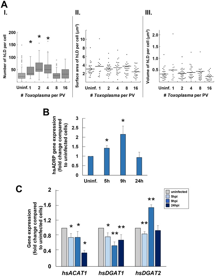
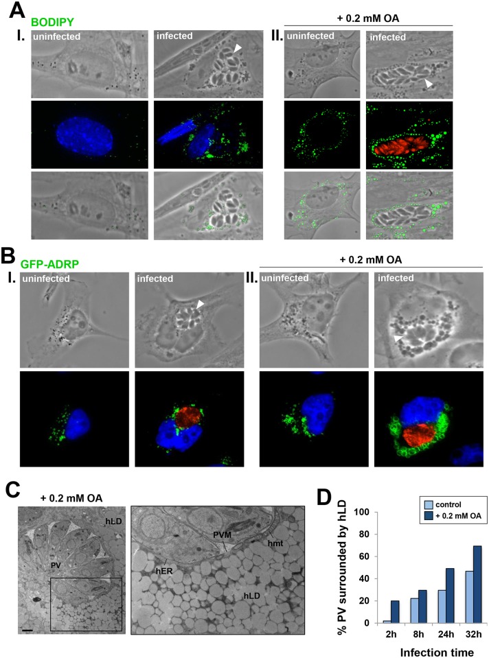
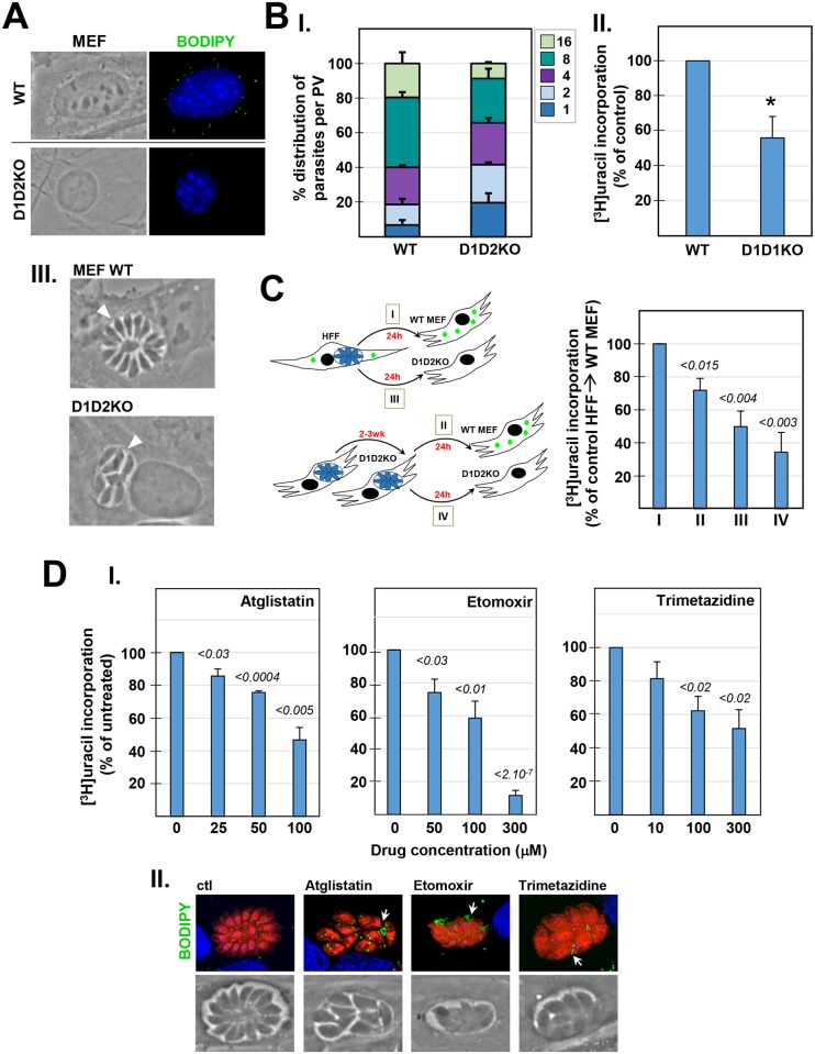
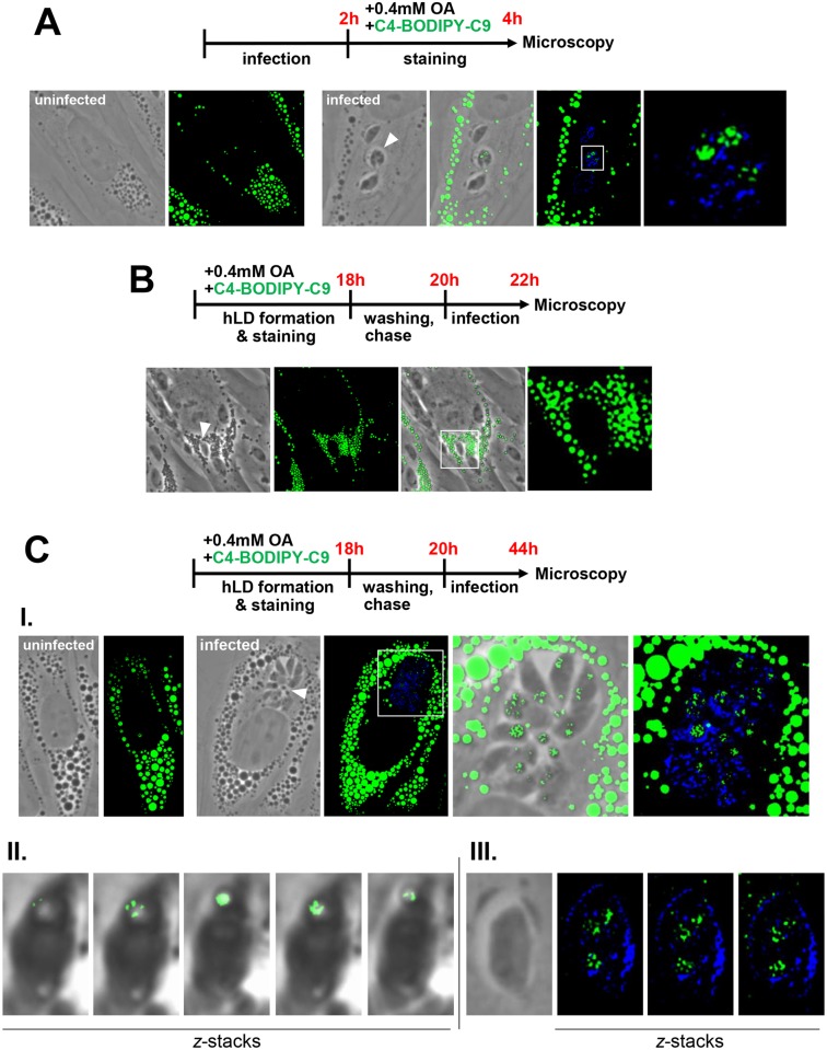
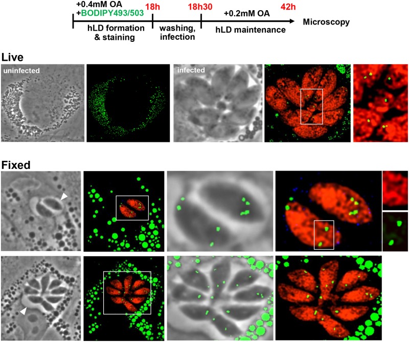
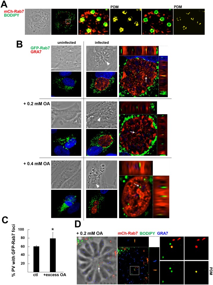
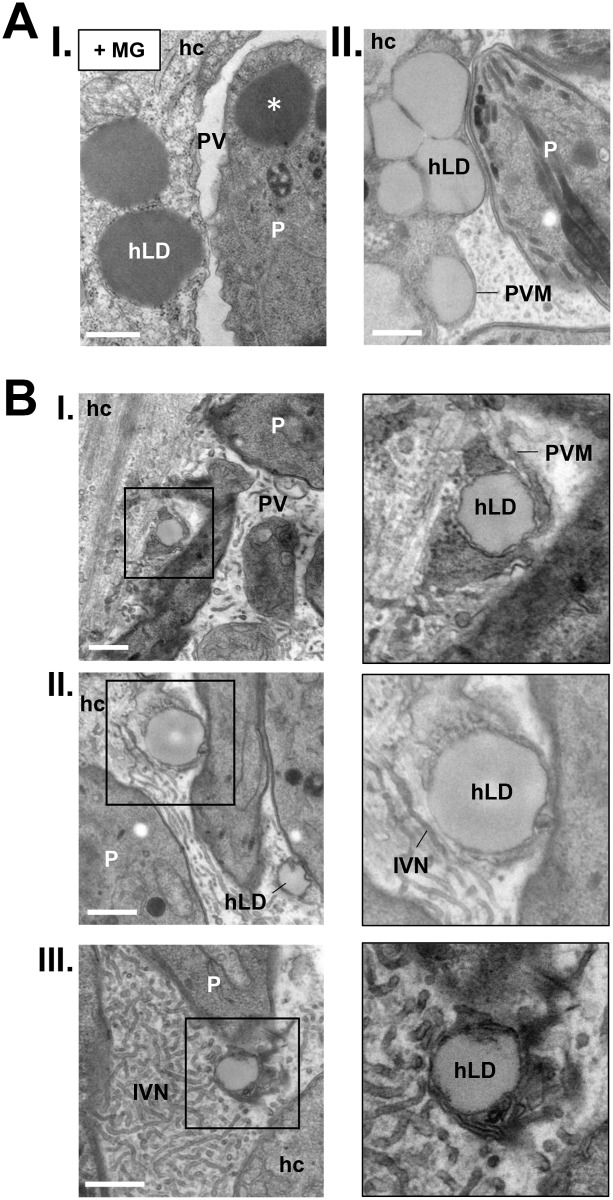
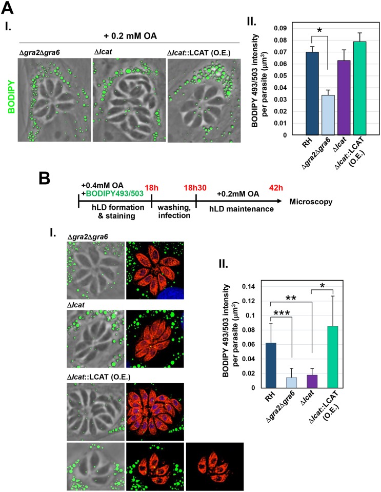
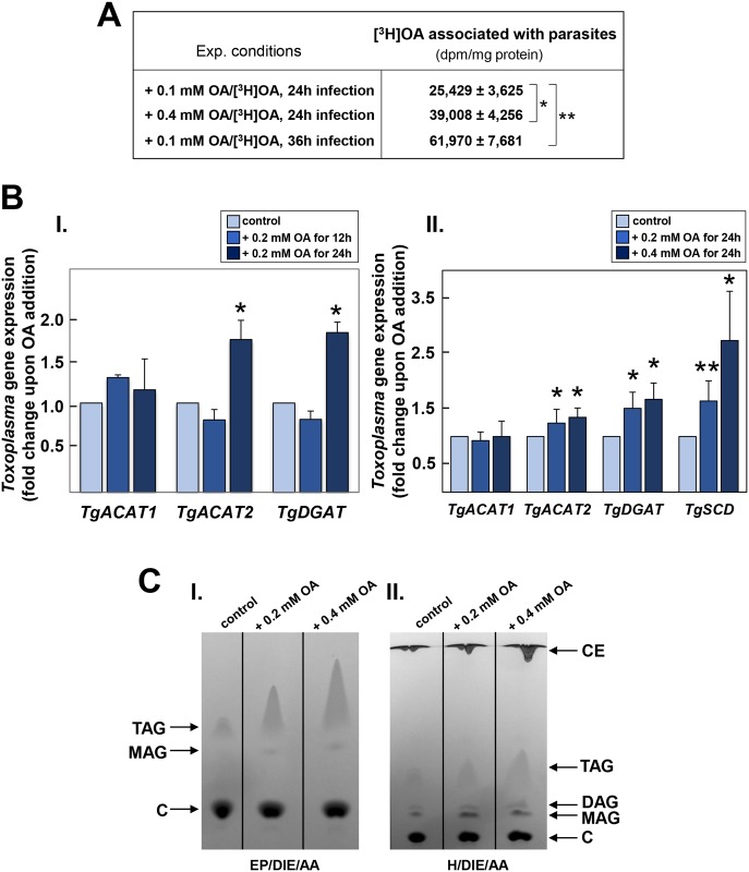
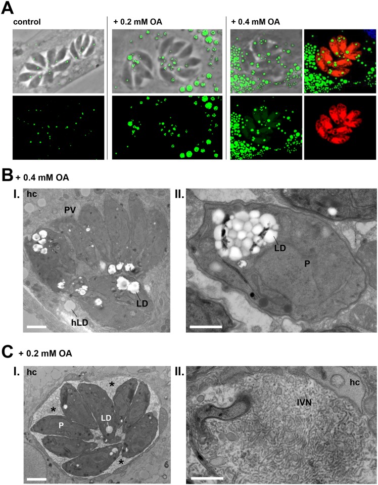
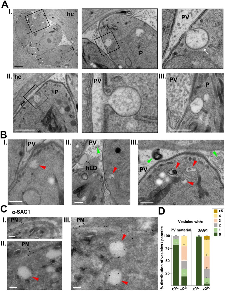
Toxoplasma is an obligate intracellular parasite that replicates in mammalian cells within a parasitophorous vacuole (PV) that does not fuse with any host organelles. One mechanism developed by the parasite for nutrient acquisition is the attraction of host organelles to the PV. Here, we examined the exploitation of host lipid droplets (LD), ubiquitous fat storage organelles, by Toxoplasma. We show that Toxoplasma replication is reduced in host cells that are depleted of LD, or impaired in TAG lipolysis or fatty acid catabolism. In infected cells, the number of host LD and the expression of host LD-associated genes (ADRP, DGAT2), progressively increase until the onset of parasite replication. Throughout infection, the PV are surrounded by host LD. Toxoplasma is capable of accessing lipids stored in host LD and incorporates these lipids into its own membranes and LD. Exogenous addition of oleic acid stimulates LD biogenesis in the host cell and results in the overaccumulation of neutral lipids in very large LD inside the parasite. To access LD-derived lipids, Toxoplasma intercepts and internalizes within the PV host LD, some of which remaining associated with Rab7, which become wrapped by an intravacuolar network of membranes (IVN). Mutant parasites impaired in IVN formation display diminished capacity of lipid uptake from host LD. Moreover, parasites lacking an IVN-localized phospholipase A2 are less proficient in salvaging lipids from host LD in the PV, suggesting a major contribution of the IVN for host LD processing in the PV and, thus lipid content release. Interestingly, gavage of parasites with lipids unveils, for the first time, the presence in Toxoplasma of endocytic-like structures containing lipidic material originating from the PV lumen. This study highlights the reliance of Toxoplasma on host LD for its intracellular development and the parasite's capability in scavenging neutral lipids from host LD.


Conflict of interest statement

The authors have declared that no competing interests exist.

Figures

Fig 1
Fig 1. Influence of Toxoplasma infection on host LD number and composition.
A. Properties of host LD in infected HFF. Determination, using Volocity software, of the host LD (hLD) number (in I), surface area (in II) and volume (inIII) throughout infection. Data were acquired from 2 independent experiments, and were categorized by the number of parasites per PV (> 30 PV counted per experiment for each sample). In panels II and III, the values of host LD surface area and volume, expressed in μm2 and μm3 respectively, represent the mean surface area and volume of host LD in individual cells, grouped together (n < 30 cells per replicate experiment), of which there were no statistical differences. Significant differences were observed for host LD number, which increased in cells containing PV with a single, 2 and 4 parasites, as compared to uninfected cells (*p <0.05). B. Human Adipose Differentiation-Related Protein (hsADRP) gene expression in infected HFF. Real-time PCR analysis of hsADRP transcripts throughout infection. Data are means ± SD of 3 separate assays performed in biological triplicates, showing significant increase of transcript levels of hsADRP at early time points of infection compared to uninfected cells (p <0.001) C. Human Acetyl-Coenzyme A Acetyltransferase 1 (hsACAT1), Diacylglycerol O-Acyltransferase 1 (hsDGAT1) and Diacylglycerol O-Acyltransferase 2 (hsDGAT2) gene expression in infected HFF. Real-time PCR analysis of hsACAT1, hsDGAT1 and hsDGAT2 throughout Toxoplasma infection. Data are means ± SD of 3 separate assays performed with biological triplicates, showing significant changes in transcript levels of the three genes early in infection compared to uninfected cells (*p <0.0003; **p <0.0001).
Fig 2
Fig 2. Influence of Toxoplasma infection on host LD distribution.
A. Fluorescence microscopy of HFF infected with RFP-expressing Toxoplasma or WT for 24 h. Host LD were identified by staining with BODIPY 493/503 (green) and 4-,6-diamidino-2-phenylindole (DAPI, blue, nucleus). Uninfected and infected cells were incubated either under control conditions (corresponding to culture medium supplemented with 10% FBS that contains on average 3 μg/ml OA; in I) or with 0.2 mM OA added to the medium (corresponding to a ~20-fold excess of OA; in II). Images show host LD gathering around each PV, which was more pronounced upon OA addition. Arrowheads pinpoint PV. B. Fluorescence microscopy of GFP-ADRP-expressing HeLa cells infected with Toxoplasma for 24 h. The PV (arrowheads) were immunostained for GRA7 (red) in cells incubated with (in I) or without OA (in II). GFP-labeled host LD encircle the PV. In blue, DAPI. C. EM of HFF infected with Toxoplasma for 24 h and incubated with 0.2 mM OA during infection, confirming the amassing of host LD at the PV membrane (PVM). Host ER (hER) and mitochondria (hmt) remained associated with the vacuolar membrane (inset). Scale bar, 1 μm. D. Quantification of the percent of the PV population surrounded by host LD during infection, with or without exogenous 0.2 mM OA. GFP-ADRP-expressing HeLa cells were infected with Toxoplasma at the indicated times. PV were scored as LD-associated if > 70% of the total host LD population was grouped around the PV.
Fig 3
Fig 3. Influence of host LD on Toxoplasma intracellular development.
A. Fluorescence microscopy of uninfected MEF, either WT or lacking DGAT1 and DGAT2 (D1D2KO) stained with BODIPY493/503 (green) and DAPI (blue, nucleus), showing very few, tiny LD in the mutant D1D2KO MEF. B. Assessment of parasite development in WT and D1D2KO MEF. The number of parasites per PV in WT and D1D2KO MEF infected for 24 h were counted (panel I). Data are means ± SD of 3 independent experiments. PV sizes were statistically different between the WT and D1D2KO conditions (Chi-squared, p <0.0001, all 3 biological replicates significant). Panel II: [3H]uracil incorporation assays to quantify parasite replication 24 h p.i. Data are percentages ± SD relative to MEF WT controls (set as 100%) of 4 independent assays performed in triplicate assays (*p <0.0008, Student’s t test). Absolute values in cpm for each experiment are shown in S2 Fig. Panel III shows phase images of representative PV in WT and D1D2KO MEF, showing no difference in parasite morphology between the 2 conditions. C. Assessment of parasite development following serial passage in D1D2KO MEF. Four experimental conditions were designed as depicted on the schema: Toxoplasma from HFF were transferred to WT MET (condition I) or D1D2KO (condition III) for 24 h before [3H]uracil incorporation assays. Toxoplasma passaged in D1D2KO (P8, P9, P10, or P12) were transferred to WT MEF (condition II) or D1D2KO (condition IV) for 24 h before uracil incorporation assays. Each passage corresponds to a separate biological assay. Data are percentages ± SEM relative to condition I (set as 100%) of 4 biological experiments performed in triplicate assays. Absolute values in cpm (means ± SD) are shown in S2 Fig. Significant differences in replication rates between condition I and other conditions were calculated using the Student’s t test. D. Effect of host ATGL, CPT-1 and 3-KAT inhibition on Toxoplasma growth. Panel I: Toxoplasma-infected HFF were incubated 24 h in the presence of the indicated concentrations of Atglistatin or Etomoxir (both DMSO control), or Trimetazidine (PBS control) before [3H]uracil incorporation assays. Data are percentages ± SEM relative to controls (set as 100%) of 3 or 4 biological experiments in triplicates. Significant differences in replication rates between the treated and control samples were calculated with the Student’s t test. Absolute values in cpm (means ± SD) are shown in S2 Fig. Panel II: fluorescence microscopy after 24 h of RFP-Toxoplasma-infected HFF stained with BODIPY 493/503 (green) and DAPI (blue), showing representative PV following exposure to Atglistatin (50 μM), Etomoxir (100 μM) or Trimetazidine (100 μM). Smaller PV and lipid deposits in the vacuolar space were observed, compared to untreated parasites.
Fig 4
Fig 4. Detection in Toxoplasma of the fatty acid C4-BODIPY-C9 present from host LD.
A-C. Fluorescence microscopy of uninfected or infected HFF in the presence of 0.4 mM OA and the free fatty acid C4-BODIPY-C9 (10 μM). Schemas outlining the experimental protocols are shown at the top of each panel. In A, C4-BODIPY-C9 (green) was co-administrated with OA for 2 h before fixation and immunolabeling of the PV with anti-GRA7 antibodies (blue), resulting in C4-BODIPY-C9 straining on the parasite. In B and C, C4-BODIPY-C9 (green) was co-administrated with OA for 18 h prior to infection, allowing the storage of C4-BODIPY-C9 in host LD (hLD). After washing and C4-BODIPY-C9 chase, cells were infected for 2 h (B) or 24 h (C). C4-BODIPY-C9 association with the parasite was apparent at 24 h (C, panel I). Panel II in C shows z-stacks of egressing parasite containing C4-BODIPY-C9 on LD as observed by live fluorescence microscopy. Panel III in C shows z-stacks of intracellular parasite stained for GRA7 (blue) and containing C4-BODIPY-C9 on cytoplasmic membranes by IFA.
Fig 5
Fig 5. Detection in Toxoplasma of BODIPY 493/503 originated from host LD.
Fluorescence microscopy of uninfected or infected HFF with RFP-expressing parasites in the presence of 0.4 mM OA and BODIPY 493/503 (10 μM). Schema outlining the experimental protocols is shown at the top. BODIPY 493/503 (green) was co-administrated with OA for 18 h prior to infection, allowing the storage of BODIPY 493/503 in host LD (hLD). After washing and infection in the presence of 0.2 mM OA, to maintain host LD, for ~24 h, cells were observed by live microscopy (upper panel) or fixed for IFA using anti-GRA7 antibodies (in blue; lower panel). In both cases, the BODIPY 493/503 signal was observed within the parasite both in small and large PV (arrowheads).
Fig 6
Fig 6. Detection of host Rab7-associated structures in the PV.
A. Fluorescence microscopy of HFF expressing mCherry-Rab7. mCherry Rab7-expressing HFF incubated with 0.2 mM OA for 24 h were fixed and stained with BODIPY 493/503. A phase and a maximum projection (extended depth of field) image displaying the merge of BODIPY 493/503 (green) and mCherry Rab7 (red) are shown. A digital magnification of the boxed area is shown as two individual z-slice images displaying the merge of BODIPY 493/503(green) and mCherry Rab7 (red) plus the positive PDM, showing a subset of host LD containing Rab7 on their surface. B. Fluorescence microscopy of uninfected or 24 h-Toxoplasma-infected HFF expressing GFP-Rab7 grown without OA, with 0.2 or 0.4 mM OA. Cells were fixed and stained with antibodies for GRA7 (red; PV) and DAPI (blue; nucleus). Arrowheads pinpoint PV on phase images. The distribution of GFP-Rab7-positive vesicles (green) is shown in both uninfected and infected cells. A digital magnification of the Toxoplasma PV is shown in an orthogonal view of the z-stack to highlight the localization of host-derived GFP-Rab7 vesicles inside the PV (arrows). C. Quantification of the percentage of PV containing GFP-Rab7-associated structures in the PV lumen. Infected HFF expressing GFP-Rab7 were incubated in the absence of added OA (control) or with excess OA (0.2 or 0.4 mM) as described in B. The number of PV containing GFP-Rab7 structures was determined by analysis of microscopy images of optical z-stacks. Data are mean values ± SD (n = 3) (PV > 20 in each experiment). Values are statistically significant between PV exposed to OA (0.2 or 0.4 mM) and without OA as control (Chi-squared test, *p <0.0129). D. Fluorescence microscopy of mCherry-Rab7 expressing HFF infected with Toxoplasma for 24 h, and stained with BODIPY 493/503 (green) and GRA7 (blue). A phase and orthogonal view of a fluorescent z-stack are shown. A digital magnification of the boxed region displayed as individual z-slices showing the BODIPY 493/503 channel (green), the mCherry-Rab7 channel (red), a merged image and positive PDM. These images illustrate the co-staining of intravacuolar structures for mCherry-Rab7 and BODIPY 493/503 (arrow).
Fig 7
Fig 7. Ultrastructural evidence of host LD projection and trapping into the PV.
A-B. Transmission EM of Toxoplasma-infected HFF for 24 h. In A, panel I: infected cells were fixed in the presence of malachite green (MG) to facilitate the identification of LD while in panel II, infected cells were incubated with 0.2 mM OA to increase LD number. In both situations, host LD (hLD) were observed protruding from the host cytoplasm into the PV lumen. The asterisk points to parasite LD. In B, infected cells were loaded with 0.2 mM OA and host LD were observed in the vacuolar space, wrapped by IVN tubules (panels I-III). hc, host cell; P, parasite; PVM, PV membrane. All scale bars, 0.5 μm.
Fig 8
Fig 8. Detection of BODIPY 493/503 originated from host LD in Toxoplasma mutants.
A. Fluorescence microscopy of HFF infected with Δgra2Δgra6, Δlcat and Δlcat::LCAT overexpressors (O.E.) for 24 h with 0.2 mM OA added to the medium. Host LD were identified by staining with BODIPY 493/503 (green) overlaid on phase contrast as shown in panel I. Panel II: the basal LD volume of the parasites was calculated by measuring the intensity of BODIPY 493/503 within individual PV, and normalized to parasite number, without the addition of OA (means ± SD of triplicate experiments, n > 400 parasites per experimental replicate and mutant (t-test: *p <0.01). B. Fluorescence microscopy of infected HFF with Δgra2Δgra6, Δlcat and Δlcat::LCAT O.E. parasites in the presence of 0.4 mM OA and BODIPY 493/503 (10 μM). Schema outlining the experimental protocols is shown at the top. BODIPY 493/503 (green) was co-administrated with OA for 18 h prior to infection, allowing the storage of BODIPY 493/503 in host LD (hLD). After washing and infection in the presence of 0.2 mM OA, to maintain host LD for 24 h, cells were fixed for IFA using antibodies against anti-Hsp70/aldolase antibodies to stain the parasite cytosol (in red). Panel I shows representative PV for Δgra2Δgra6 and Δlcat parasites for which the BODIPY 493/503 signal was weak as compared to Δlcat::LCAT overexpressors (large and small PV). Panel II: the average LD volume of the parasites was calculated using Volocity to determine the averaged BODIPY 493/503 intensity, thus volume, within parasites. Data are means of means of 3 independent experiments, n > 400 parasites per experimental replicate and mutant (t-test: ***p <0.008; **p <0.02; *p <0.04).
Fig 9
Fig 9. Influence of excess oleate on parasite neutral lipid synthesis.
A. Uptake assays of radioactive OA by Toxoplasma. HFF were preincubated with 0.1 or 0.4 mM OA containing [3H]OA for 24 h, thoroughly washed and infected with Toxoplasma for 24 h or 36 h before chemically inducing parasite egress. Parasites were collected and purified to monitor their radioactivity by counting. Data of [3H]OA associated with the parasites expressed in dpm/mg cell protein, are means ± SD of 3 independent assays made in triplicates. *p <0.05; **p <0.001. B. Comparison of Toxoplasma ACAT and DGAT gene expression under conditions of excess OA or without added OA (control). Real-time PCR analysis of TgACAT1, TgACAT2, TgDGAT and TgSDC transcripts in parasites infecting cells exposed to 0.2 mM OA for 12 h or 24 h (panel I), or to 0.2 or 0.4 mM for 24 h (panel II). Data are means ± SD of 3 independent assays performed in triplicates. Transcripts levels of TgACAT2 and TgDGAT were significantly increased in the presence of excess OA compared to control conditions in the absence of OA (*p <0.004; **p <0.0001). C. TLC analysis of neutral lipid fractions of cellular lipid extracts from intracellular Toxoplasma exposed to excess OA. Neutral lipids were separated on plates using as solvent EP/DIE/AA (panel I) to resolve TAG and MAG, or H/DIE/AA (panel II) to separate CE and DAG. CE and species of acylglycerols were more abundantly produced under conditions of excess OA vs. no OA added.
Fig 10
Fig 10. Influence of excess oleate on parasite neutral lipid stores and IVN size.
A. Fluorescence microscopy of RFP-expressing Toxoplasma-infected HFF. Infected cells were cultivated without OA (control), or with 0.2 mM OA for 24 h or 0.4 mM OA for 40 h before staining with BODIPY 493/503 (green). Large, fluorescent neutral lipid deposits inside Toxoplasma were observed in the presence of 0.4 mM OA. These deposits are confirmed by the presence of cytoplasmic exclusion zones of the RFP fluorescence in the parasite. B. Transmission EM images of HFF infected for 24 h in the presence of 0.4 mM OA, showing many parasite LD with some as large as host LD (hLD; panel I) and others clustered inside the parasite (panel II). Scale bars, 0.5 μm. C. Transmission EM images of a Toxoplasma PV inside HFF infected for 24 h in the presence of 0.2 mM OA. Panel I (asterisks) shows the expansion of the IVN throughout the vacuolar space, taking up all available space between parasites while panel II shows a magnified view focusing on the abundance of long, intertwined tubules. P, parasite; hc, host cell. Scale bars, 0.3 μm.
Fig 11
Fig 11. Ultrastructural detection of endocytic-like structures in Toxoplasma.
A-B. Transmission EM of parasites in HFF incubated with 0.2 mM OA for 24 h. In A, panels I-III show three plasma membrane invaginations decorated with a cytoplasmic coat (arrow in I) and whose content displays resemblance with the material present in the PV lumen, suggestive of endocytic events. P, parasite; hc, host cell. Scale bars, 0.3 μm. In B, panels I-III illustrate intraparasitic vesicles containing osmiophilic material similar to that present in the PV lumen close to the parasite (red arrowheads). C. ImmunoEM for SAG1 detection, on parasites in HFF incubated with 0.2 mM OA for 24 h for SAG1 detection. Anti-SAG1-gold particles were observed on the limiting membrane of intracytoplasmic vesicles (red arrowheads). This antibody recognizes SAG1 epitopes on the extracellular moiety of SAG1 (panel I), making the SAG1 staining distributed on the internal leaflet of the organellar membrane, as expected for an endocytic event (panels II and III). PM, parasite plasma membrane. D. Quantification of intracytoplasmic vesicles either containing PV material or SAG1 staining in control and OA-loaded parasites. Data are means ± SD of 65 to 80 PV observed. All scale bars in A-C, 0.1 μm.

References

    1. Bartz R, Li WH, Venables B, Zehmer JK, Roth MR, Welti R, et al. (2007) Lipidomics reveals that adiposomes store ether lipids and mediate phospholipid traffic. J Lipid Res 48: 837–847. 10.1194/jlr.M600413-JLR200 - DOI - PubMed
    1. Brasaemle DL (2007) The perilipin family of structural lipid droplet proteins: stabilization of lipid droplets and control of lipolysis. J Lipid Res 48: 2547–2559. 10.1194/jlr.R700014-JLR200 - DOI - PubMed
    1. Kuerschner L, Moessinger C, Thiele C (2008) Imaging of lipid biosynthesis: how a neutral lipid enters lipid droplets. Traffic 9: 338–352. 10.1111/j.1600-0854.2007.00689.x - DOI - PubMed
    1. Stone SS, Levin MC, Zhou P, Han J, Walther TC, Farese RV Jr (2009) The endoplasmic reticulum enzyme, DGAT2, is found in mitochondria-associated membranes and has a mitochondrial targeting signal that promotes its association with mitochondria. J Biol Chem 284: 5352–5361. 10.1074/jbc.M805768200 - DOI - PMC - PubMed
    1. Fujimoto T, Parton RG (2011) Not just fat: the structure and function of the lipid droplet. Cold Spring Harb Perspect Biol 3: a004838 10.1101/cshperspect.a004838 - DOI - PMC - PubMed