

Consensus on Diagnostic Criteria of Idiopathic Orbital Inflammation Using a Modified Delphi Approach
- PMID: 28570731
- PMCID: PMC5710199
- DOI: 10.1001/jamaophthalmol.2017.1581
Consensus on Diagnostic Criteria of Idiopathic Orbital Inflammation Using a Modified Delphi Approach
Abstract
Importance: Current practice to diagnose idiopathic orbital inflammation (IOI) is inconsistent, leading to frequent misdiagnosis of other orbital entities, including cancer. By specifying criteria, diagnosis of orbital inflammation will be improved.
Objective: To define a set of criteria specific for the diagnosis of IOI.
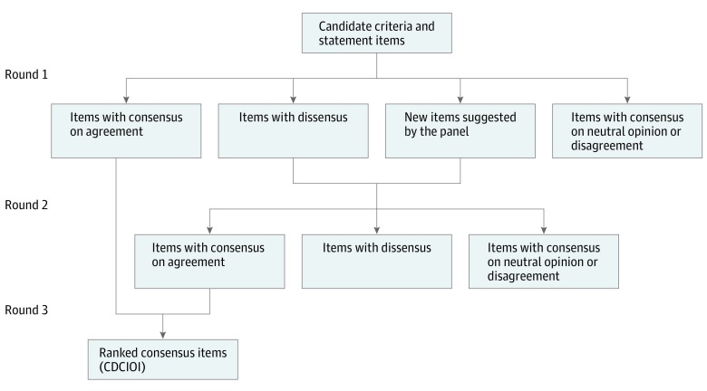
Design, setting, and participants: A 3-round modified Delphi process with an expert panel was conducted from June 8, 2015, to January 25, 2016. Fifty-three orbital scientist experts, identified through membership in the Orbital Society, were invited to participate in on online survey and they scored, using 5-point Likert scales, items that are eligible as diagnostic criteria from the literature and from personal experience. The items were clustered around the anatomic subtypes of IOI: idiopathic dacryoadenitis and idiopathic orbital fat inflammation (2 nonmyositic IOIs), and idiopathic orbital myositis (myositic IOI). Items with dissensus were rescored in the second round, and all items with consensus (median, ≥4; interquartile range, ≤1) were ranked by importance in the third round.
Main outcomes and measures: Consensus on items to be included in the criteria.
Results: Of the 53 experts invited to participate, a multinational panel of 35 (66%) individuals with a mean (SD) years of experience of 31 (11) years were included. Consensus was achieved on 7 of 14 clinical and radiologic items and 5 of 7 pathologic items related to diagnosis of nonmyositic IOI, and 11 of 14 clinical and radiologic items and 1 of 5 pathologic items for myositic IOI. There was agreement among panelists to focus on surgical tissue biopsy results in the diagnosis of nonmyositic IOI and on a trial with systemic corticosteroids in myositic IOI. Panelists agreed that a maximum number of 30 IgG4-positive plasma cells per high-power field in the orbital tissue is compatible with the diagnosis of IOI.
Conclusions and relevance: An international panel of experts endorsed consensus diagnostic criteria of IOI. These criteria define a level of exclusion suggested for diagnosis and include tissue biopsy for lesions not confined to the extraocular muscles. This consensus is a step toward developing guidelines for the management of IOI, which needs to be followed by validation studies of the criteria.
Conflict of interest statement
Figures

Comment in
-
[Orbital pseudotumor].Ophthalmologe. 2021 Aug;118(8):774-776. doi: 10.1007/s00347-021-01437-x. Epub 2021 Aug 5. Ophthalmologe. 2021. PMID: 34351446 German. No abstract available.
References
-
- Mombaerts I, Goldschmeding R, Schlingemann RO, Koornneef L. What is orbital pseudotumor? Surv Ophthalmol. 1996;41(1):66-78. - PubMed
-
- Kahana A, Elner VM. The meaning of diagnoses in orbital disease. Ophthal Plast Reconstr Surg. 2013;29(5):347-348. - PubMed
-
- Ben Simon GJ, Yoon MK, Atul J, Nakra T, McCann JD, Goldberg RA. Clinical manifestations of orbital mass lesions at the Jules Stein Eye Institute, 1999-2003. Ophthalmic Surg Lasers Imaging. 2006;37(1):25-32. - PubMed
-
- Harris GJ. Idiopathic orbital inflammation: a pathogenetic construct and treatment strategy: the 2005 ASOPRS Foundation Lecture. Ophthal Plast Reconstr Surg. 2006;22(2):79-86. - PubMed
-
- Dagi Glass LR, Freitag SK. Orbital inflammation: corticosteroids first. Surv Ophthalmol. 2016;61(5):670-673. - PubMed
MeSH terms
LinkOut - more resources
Full Text Sources
Other Literature Sources
