

Acute onset of paraganglioma of filum terminale: A case report and surgical treatment
- PMID: 28570879
- PMCID: PMC5453863
- DOI: 10.1016/j.ijscr.2017.05.016
Acute onset of paraganglioma of filum terminale: A case report and surgical treatment
Abstract
Introduction: Paragangliomas of filum terminale are rare benign tumors, arising from the adrenal medulla or extra-adrenal paraganglia. These lesions usually present with chronic back pain and radiculopathy and only two cases of acute neurological deficit have been reported in literature.
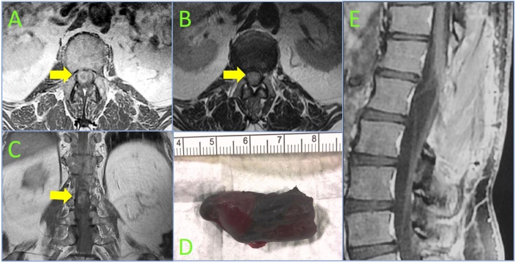
Presentation of case: A case with an acute paraplegia and cauda equina syndrome due to an hemorrhagic paraganglioma of the filum terminale is described. Magnetic resonance imaging showed an intradural tumor extending from L1 to L2 compressing the cauda equina, with an intralesional and intradural bleed. An emergent laminectomy with total removal of the tumor was performed allowing a post-operative partial sensory recovery. Histopathological examination diagnosed paraganglioma.
Discussion: Paragangliomas are solid, slow growing tumors arising from specialized neural crest cells, mostly occurring in the head and neck and rarely in cauda equina or filum terminale. MRI is gold standard radiological for diagnosis and follow-up of these lesions. They have no pathognomonic radiological and clinical features and are frequently misdiagnosed as other spinal lesions. No significant correlation was observed between the duration of symptoms and tumor dimension. Acute presentation is unusual and emergent surgical treatment is fondamental. The outcome is very good after complete excision and radiotherapical treatment is recommended after an incomplete resection.
Conclusion: Early radiological assessment and timely surgery are mandatory to avoid progressive neurological deficits in case of acute clinical manifestation of paraganglioma of filum terminale.
Keywords: Case report; Cauda equina; Filum terminale; Paraganglioma; Spinal tumor.
Copyright © 2017 The Author(s). Published by Elsevier Ltd.. All rights reserved.
Figures

References
-
- Dillard-Cannon E., Atsina K.B., Ghobrial G., Gnass E., Curtis M.T., Heller J. Lumbar paraganglioma. J. Clin. Neurosci. 2016;30:149–151. - PubMed
-
- Gutenberg A., Wegner C., Pilgram-Pastor S.M., Gunawan B., Rohde V., Giese A. Paraganglioma of the filum terminale: review and report of the first case analyzed by CGH. Clin. Neuropathol. 2010;29:227–232. - PubMed
-
- Agha R.A., Fowler A.J., Saeta A., Barai I., Rajmohan S., Orgill D.P., Group S. The SCARE statement: consensus-based surgical case report guidelines. Int. J. Surg. 2016;34:180–186. - PubMed
-
- Lerman R.I., Kaplan E.S., Daman L. Ganglioneuroma-paraganglioma of the intradural filum terminale. Case report. J. Neurosurg. 1972;36:652–658. - PubMed
-
- Masuoka J., Brandner S., Paulus W., Soffer D., Vital A., Chimelli L., Jouvet A., Yonekawa Y., Kleihues P., Ohgaki H. Germline SDHD mutation in paraganglioma of the spinal cord. Oncogene. 2001;20:5084–5086. - PubMed
LinkOut - more resources
Full Text Sources
Other Literature Sources
