Comparative Study
. 2017 Sep:43:76-86.
doi: 10.1016/j.tiv.2017.05.021. Epub 2017 May 30.

Comparison of in vitro toxicological effects of biomass smoke from different sources of animal dung

Affiliations
Comparative Study

Comparison of in vitro toxicological effects of biomass smoke from different sources of animal dung

Claire E McCarthy et al. Toxicol In Vitro. 2017 Sep.

Abstract

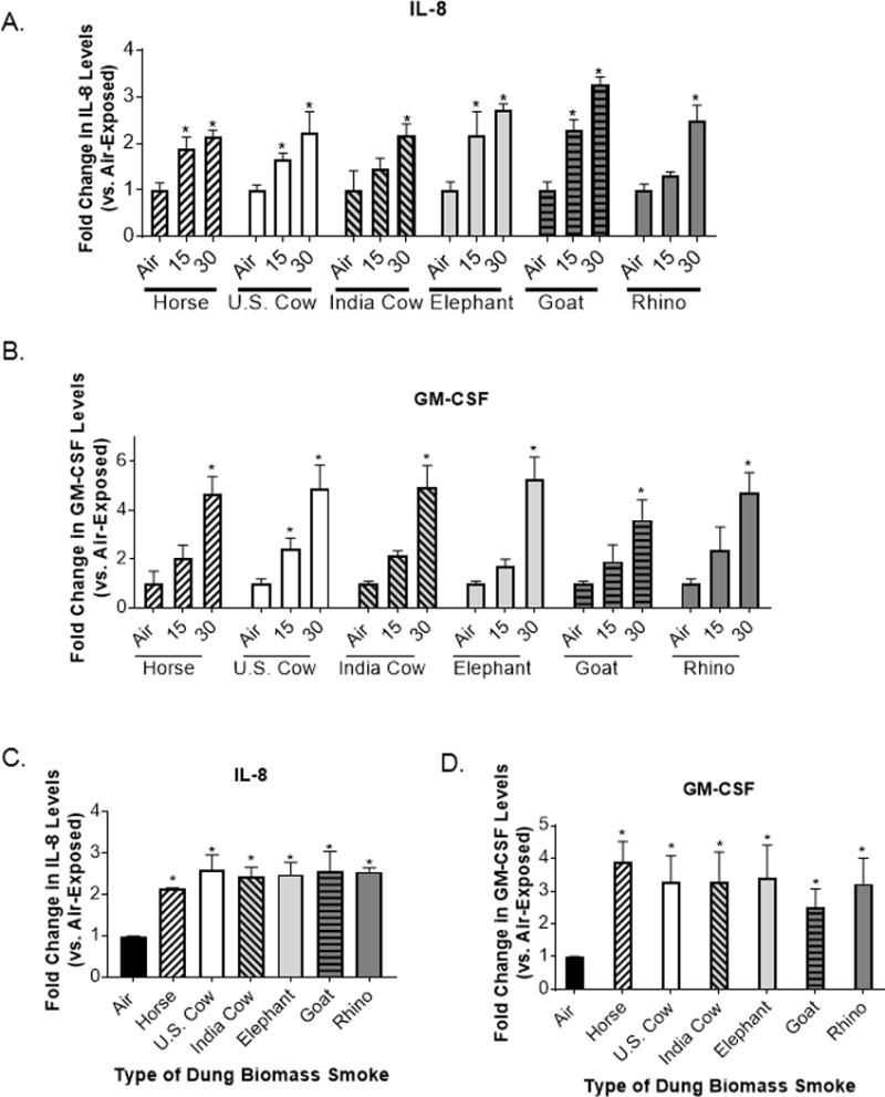
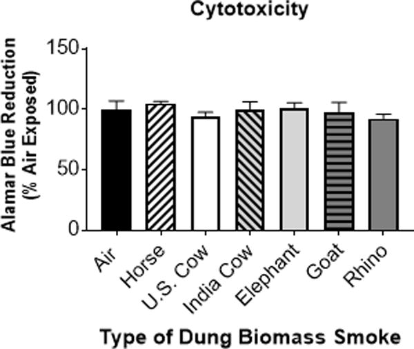
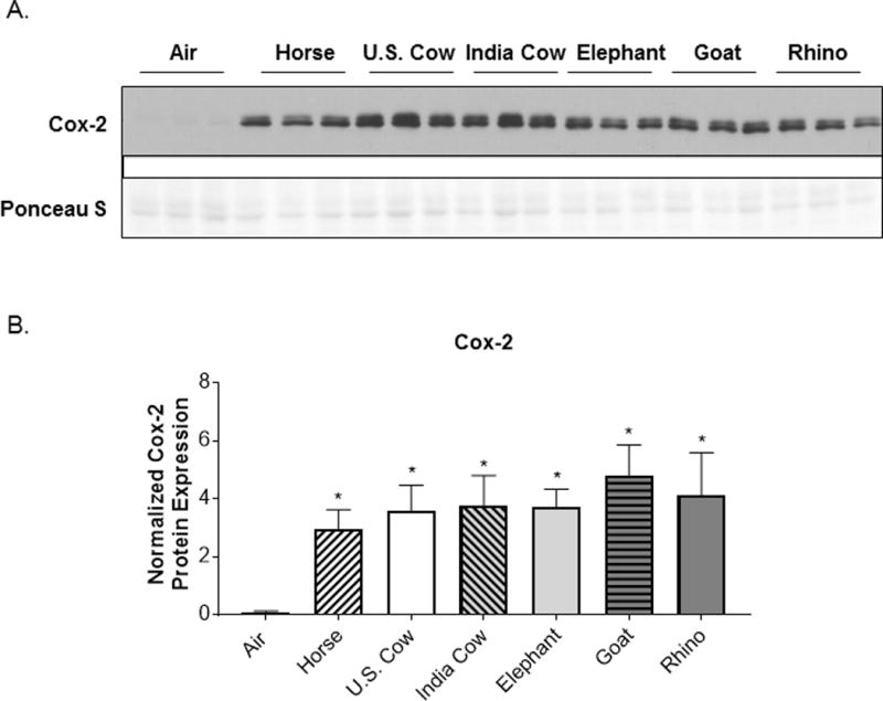
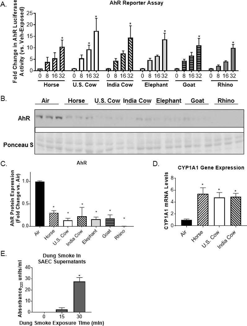
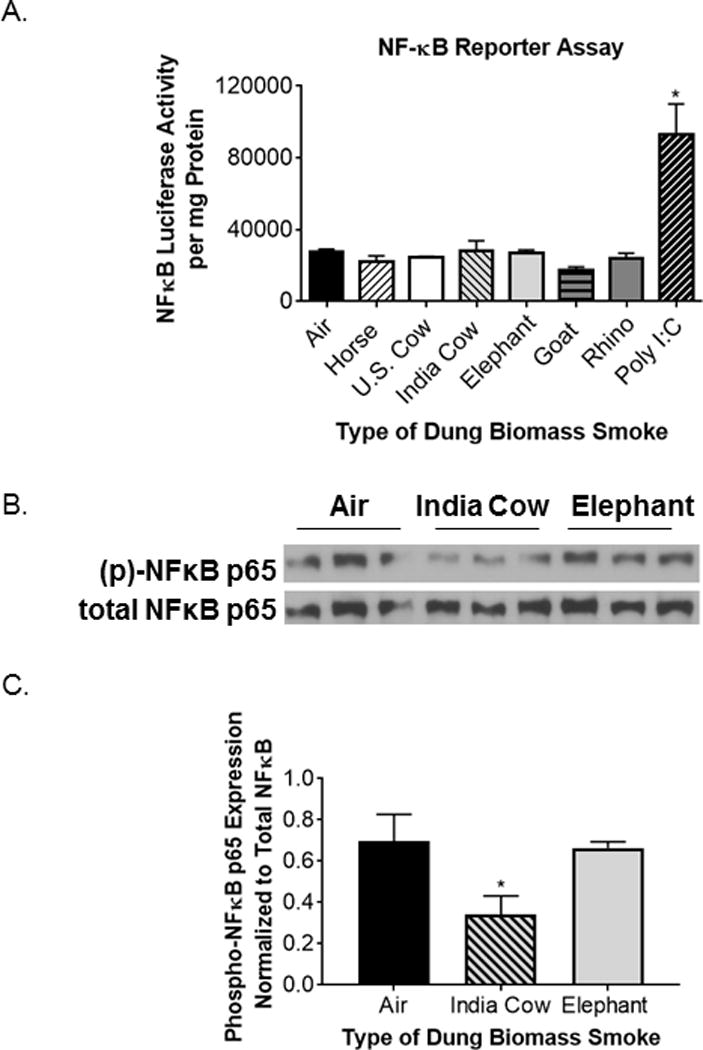
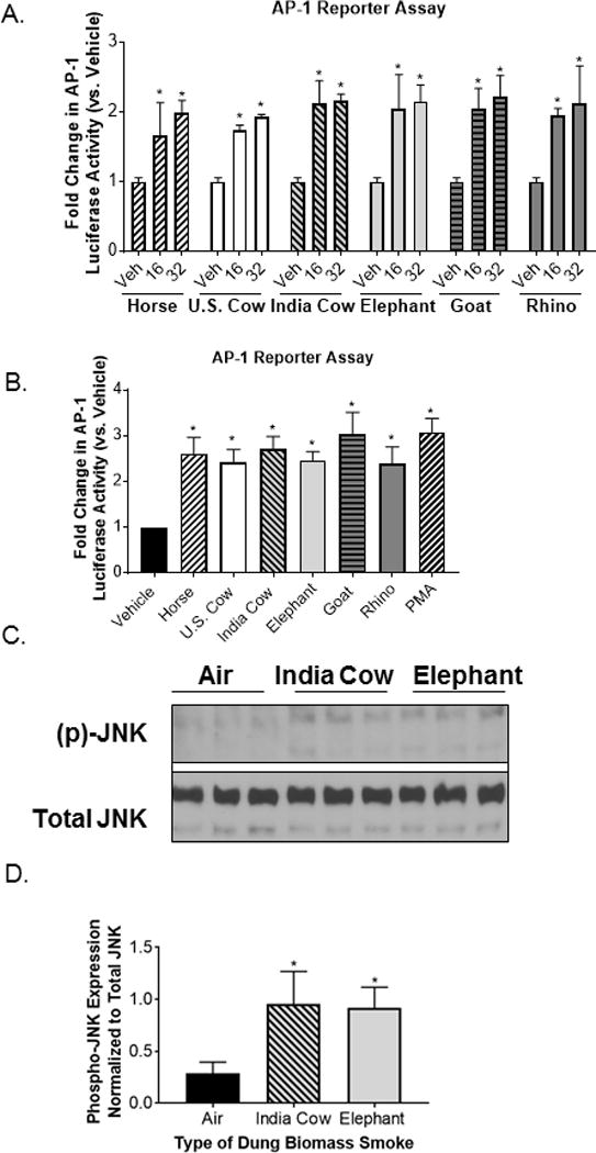
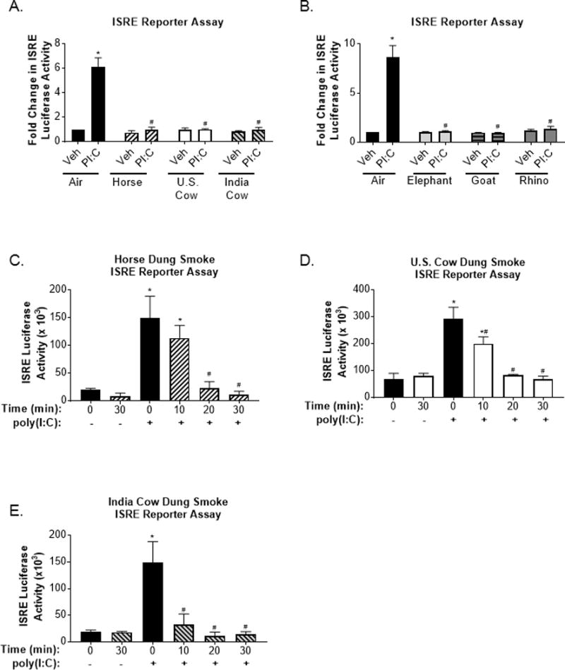
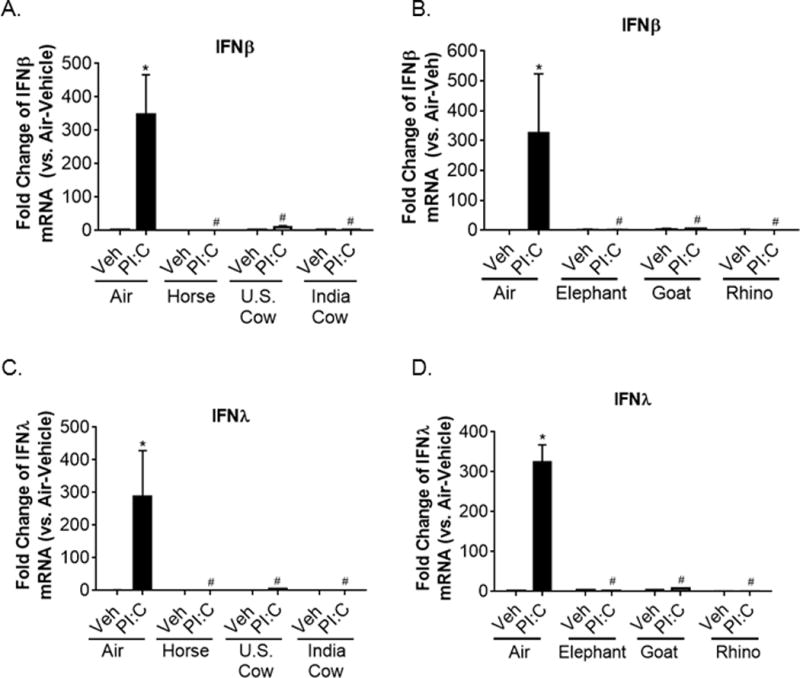
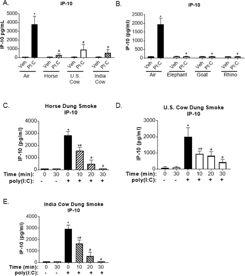
Worldwide, over 4 million premature deaths each year are attributed to the burning of biomass fuels for cooking and heating. Epidemiological studies associate household air pollution with lung diseases, including chronic obstructive pulmonary disease, lung cancer, and respiratory infections. Animal dung, a biomass fuel used by economically vulnerable populations, generates more toxic compounds per mass burned than other biomass fuels. The type of animal dung used varies widely depending on local agro-geography. There are currently neither standardized experimental systems for dung biomass smoke research nor studies assessing the health impacts of different types of dung smoke. Here, we used a novel reproducible exposure system to assess outcomes related to inflammation and respiratory infections in human airway cells exposed to six different types of dung biomass smoke. We report that dung biomass smoke, regardless of species, is pro-inflammatory and activates the aryl hydrocarbon receptor and JNK transcription factors; however, dung smoke also suppresses interferon responses after a challenge with a viral mimetic. These effects are consistent with epidemiological data, and suggest a mechanism by which the combustion of animal dung can directly cause lung diseases, promote increased susceptibility to infection, and contribute to the global health problem of household air pollution.

Keywords: Biomass smoke; Household air pollution; Respiratory toxicology.


Figures

Figure 1
Figure 1
Six different types of dung biomass smoke induce pro-inflammatory cytokine production in SAECs. SAECs were exposed to air or dung smoke (horse, U.S. cow, India cow, elephant, goat, or rhinoceros (rhino)) for 15 or 30 minutes and cell supernatants were collected 24-hours post-exposure. Dose-response effects of dung biomass smoke exposure on (A) IL-8 and (B) GM-CSF production in SAECs were determined by ELISA. Data represent mean ± SD (n = 3 – replicates per exposure group from an independent experiment), *p<0.05 by two-way ANOVA (compared to air-exposed cells using Tukey’s post-hoc analysis). (C) IL-8 and (D) GM-CSF levels were measured in SAECs exposed to 30 minutes of dung smoke in multiple experiments. Data represent mean ± SEM for n=3 independent experiments with 3 replicate cultures per experiment.*p<0.05 by one-way ANOVA (compared to air-exposed cells using a Dunnett’s post-hoc analysis).
Figure 2
Figure 2
Exposure to 30 minutes of six different types of dung biomass smoke does not cause cytotoxicity in SAECs. SAECs were exposed to air or dung smoke (horse, U.S. cow, India cow, elephant, goat, or rhinoceros (rhino)) for 30 minutes. Alamar Blue reagent was added to the cells 24-hours post-exposure. After a 4-hour incubation, cell supernatants containing Alamar Blue were collected. Reduction of Alamar Blue was determined by measuring the fluorescence of the samples (excitation = 560 nm and emission = 590 nm). Data represent mean (n = 3 replicates per exposure group from an independent experiment).
Figure 3
Figure 3
Cox-2 protein expression is upregulated by six different types of dung biomass smoke. SAECs were exposed to air or dung smoke (horse, U.S. cow, India cow, elephant, goat, or rhinoceros (rhino)) for 30 minutes and cell lysates were collected 24-hours post-exposure. (A) Representative Western blot showing 3 replicates per condition of Cox-2 protein expression and Ponceau S staining in SAECs exposed to air or different types of dung smoke. (B) Densitometry of Cox-2 protein levels was performed. Data represent mean ± SD (n = 3 replicates per exposure group from 1 of 3 independent experiments), *p<0.05 by one-way ANOVA (compared to air-exposed cells using Tukey’s post-hoc analysis).
Figure 4
Figure 4
Dung smoke contains AhR ligands and activates the AhR. (A) A cell line containing an AhR luciferase reporter was treated with the indicated concentrations of dung biomass smoke extract (horse U.S. cow, India cow, elephant, goat, or rhinoceros (rhino)) for 24 hours. Data represent mean ± SEM for 3 independent experiments with 3 replicate cultures per experiment, *p<0.05 by two-way ANOVA (compared to vehicle using Tukey’s post-hoc analysis). (B) SAECs were exposed to dung biomass smoke for 30 minutes and cell lysates were collected 24 hours post-exposure. AhR was detected by western blot. (C) Densitometry of AhR protein expression was performed. Data represent mean ± SD (n = 3 replicates per exposure group from 1 of 3 independent experiments), *p<0.05 by one-way ANOVA (compared to air-exposed cells using Tukey’s post-hoc analysis). (D) SAECs were exposed to horse, U.S. cow, or Indian cow dung smoke for 30 minutes. RNA was isolated 6 hours post-exposure. (D) CYP1A1 gene expression was measured by qPCR and normalized to 18S mRNA levels. Data represent mean ± SD (n = 3 replicates per exposure group from an independent experiment), *p<0.05 by one-way ANOVA (compared to air-exposed cells using Tukey’s post-hoc analysis). (E) SAECs were exposed to dung biomass smoke for up to 30 minutes. Cell supernatants were collected 24-hours post-exposure and their optical density at 320 nm was measured. Data represent mean + SEM (n=3 – mean of 3 independent experiments), *p<0.05 by one-way ANOVA (compared to air-exposed cells using Tukey’s post-hoc analysis).
Figure 5
Figure 5
Six types of dung biomass smoke do not activate NFκB in human SAECs. (A) Luciferase activity was measured in SAECs transduced with a NFκB luciferase reporter 24 hours post-dung biomass smoke (horse, U.S. cow, India cow, elephant, goat, or rhino) exposure. Based on a previous study, polyinosinic:polycytidylic acid (poly I:C, 0.5 μg/ml) was used as a positive control. Data represent mean ± SD (n= 3 replicates per exposure group from an independent experiment), *p<0.05 by one-way ANOVA (compared to air-exposed cells). (B) SAECs were exposed to air, Indian cow dung smoke, or elephant dung smoke for 30 minutes and cell lysates were collected 30 minutes post-exposure. A representative Western blot showing 3 technical replicates per exposure group is shown. (C) Densitometry of phospho-NFκB p65 and total NFκB p65 expression was performed on 3 replicates per exposure group. Data represent mean ± SD (n = 3 replicates per exposure group from a representative experiment), *p<0.05 by one-way ANOVA (compared to air-exposed cells).
Figure 6
Figure 6
Different types of dung biomass smoke activate JNK-AP-1 in airway epithelial cells. (A) 16-HBEs were transfected with an AP-1 luciferase reporter and treated with 16 or 32 units/ml of DSE (horse, U.S. cow, India cow, elephant, goat, or rhino) for 24 hours. AP-1 luciferase activity was measured in cell lysates. Data represent mean ± SD (n = 3 replicates per exposure group from an independent experiment), *p<0.05 by two-way ANOVA (compared to vehicle (Veh)-treated cells using Tukey’s multiple comparison post-hoc analysis). (B) AP-1 luciferase activity in 16-HBEs treated with 32 units/ml of DSE. Phorbol myristate acetate (PMA, 50 nM) was included as a positive control. Data represent mean ± SEM (n = 3 – mean of 3 independent experiments), *p<0.05 by one-way ANOVA (compared to Veh-treated cells using Dunnett’s post-hoc analysis). (C) SAECs were exposed to air, Indian cow dung smoke, or elephant dung smoke (30 minutes) and cell lysates were collected 30 minutes post-exposure. A representative Western blot showing 3 technical replicates per exposure group is shown. (C) Densitometry of phospho-JNK and total JNK expression was performed on 3 replicates per exposure group. Data represent mean ± SD (n = 3 replicates per exposure group from an independent experiment), *p<0.05 by one-way ANOVA (compared to air-exposed cells using Tukey’s post-hoc analysis).
Figure 7
Figure 7
Interferon production in response to a viral-like challenge is attenuated in SAECs exposed to various types of dung biomass smoke. SAECs were exposed to (A) horse, U.S. cow, or India cow dung biomass smoke or (B) elephant, goat, or rhino dung biomass smoke for 30 minutes and subsequently treated with poly(I:C) (PI:C; 0.5 μg/ml) or a vehicle control (Veh) of SAGM. Cell supernatants were collected 24 hours post-PI:C treatment. HEK cells containing an ISRE reporter were incubated with the SAEC supernatants and media (1:1 v/v) for 24 hours to assess interferon levels. Data are expressed as mean ± SEM (n=3 – mean of independent experiments),*p<0.05 (Veh compared to PI:C) and #p<0.05 (Air compared to Dung Smoke) by two-way ANOVA using Sidak’s post-hoc analysis. Exposure-response effects on poly(I:C)-induced IFN production in SAECs exposed to whole (C) horse, (D) U.S. cow, or (E) Indian cow dung biomass smoke for 10, 20, or 30 minutes were also assessed. Data are expressed as mean ± SD (n=3 replicates per group from an independent experiment), *p<0.05 (Veh compared to PI:C) and #p<0.05 (Air compared to Dung Smoke) by two-way ANOVA using using Sidak’s and Tukey’s post-hoc analyses.
Figure 8
Figure 8
Poly(I:C)-induced type I and type III interferon gene expression is suppressed in SAECs exposed to multiple types of dung biomass smoke. SAECs were exposed to the indicated type of dung biomass smoke for 30 minutes and challenged with PI:C. RNA was isolated 6 hours post-PI:C treatment. (A-B) Interferon-β, (C-D) interferon-λ, and 18S RNA gene expression were assessed by qPCR. Data are expressed as mean ± SD (n=3 replicates per group from an independent experiment),*p<0.05 (Veh compared to PI:C) and #p<0.05 (Air compared to Dung Smoke) by two-way ANOVA using Sidak’s and Tukey’s post-hoc analyses.
Figure 9
Figure 9
Poly(I:C)-induced IP-10 expression is suppressed in SAECs exposed to multiple types of dung biomass smoke. SAECs were exposed to (A) horse, U.S. cow, or India cow dung biomass smoke or (B) elephant, goat, or rhino dung biomass smoke for 30 minutes and subsequently treated with poly(I:C) (PI:C; 0.5 μg/ml) or vehicle (Veh). Cell supernatants were collected 24 hours post-PI:C treatment and IP-10 levels were measured by ELISA. Data are expressed as mean ± SEM (n=3 – mean of independent experiments),*p<0.05 (Veh compared to PI:C) and #p<0.05 (Air compared to Dung Smoke) by two-way ANOVA using using Sidak’s and Tukey’s post-hoc analyses. Exposure-response effects on poly(I:C)-induced IP-10 production in SAECs exposed to whole (C) horse, (D) U.S. cow, or (E) Indian cow dung biomass smoke for 10, 20, or 30 minutes were determined by ELISA. Data are expressed as mean ± SD (n=3 replicates per group from an independent experiment), *p<0.05 (Veh compared to PI:C) and #p<0.05 (Air compared to Dung Smoke) by two-way ANOVA using Sidak’s and Tukey’s post-hoc analyses.

Similar articles

Cited by

References

    1. Martin WJ, 2nd, Glass RI, Balbus JM, Collins FS. Public health. A major environmental cause of death. Science (New York, NY) 2011;334(6053):180–181. - PMC - PubMed
    1. Yadama GN. Fires, fuel, and the fate of 3 billion: The state of the energy impoverished. Oxford: Oxford University Press; 2013.
    1. Martin WJ, II, Glass RI, Araj H, Balbus J, Collins FS, Curtis S, Diette GB, Elwood WN, Falk H, Hibberd PL, et al. Household air pollution in low- and middle-income countries: Health risks and research priorities. PLoS Med. 2013;10(6):e1001455. - PMC - PubMed
    1. Gordon SB, Bruce NG, Grigg J, Hibberd PL, Kurmi OP, Lam K-bH, Mortimer K, Asante KP, Balakrishnan K, Balmes J, et al. Respiratory risks from household air pollution in low and middle income countries. The Lancet Respiratory Medicine. 2014;2(10):823–860. - PMC - PubMed
    1. World Health Organization. Burden of disease from household air pollution for 2012. 2014

Publication types

LinkOut - more resources