. 2017 Jun 27;474(14):2301-2313.
doi: 10.1042/BCJ20170093.

G-protein-coupled receptor kinase-2 is a critical regulator of TNFα signaling in colon epithelial cells

Affiliations

G-protein-coupled receptor kinase-2 is a critical regulator of TNFα signaling in colon epithelial cells

Michael D Steury et al. Biochem J. .

Abstract

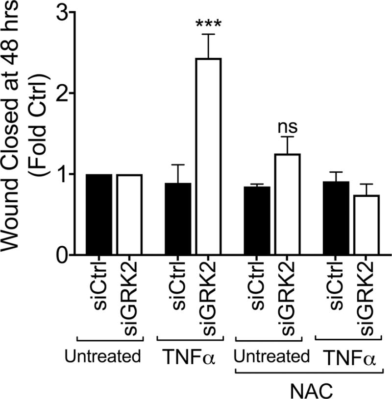
G-protein-coupled receptor kinase-2 (GRK2) belongs to the GRK family of serine/threonine protein kinases critical in the regulation of G-protein-coupled receptors. Apart from this canonical role, GRK2 is also involved in several signaling pathways via distinct intracellular interactomes. In the present study, we examined the role of GRK2 in TNFα signaling in colon epithelial cell-biological processes including wound healing, proliferation, apoptosis, and gene expression. Knockdown of GRK2 in the SW480 human colonic cells significantly enhanced TNFα-induced epithelial cell wound healing without any effect on apoptosis/proliferation. Consistent with wound-healing effects, GRK2 knockdown augmented TNFα-induced matrix metalloproteinases (MMPs) 7 and 9, as well as urokinase plasminogen activator (uPA; factors involved in cell migration and wound healing). To assess the mechanism by which GRK2 affects these physiological processes, we examined the role of GRK2 in TNFα-induced MAPK and NF-κB pathways. Our results demonstrate that while GRK2 knockdown inhibited TNFα-induced IκBα phosphorylation, activation of ERK was significantly enhanced in GRK2 knockdown cells. Our results further demonstrate that GRK2 inhibits TNFα-induced ERK activation by inhibiting generation of reactive oxygen species (ROS). Together, these data suggest that GRK2 plays a critical role in TNFα-induced wound healing by modulating MMP7 and 9 and uPA levels via the ROS-ERK pathway. Consistent with in vitro findings, GRK2 heterozygous mice exhibited enhanced intestinal wound healing. Together, our results identify a novel role for GRK2 in TNFα signaling in intestinal epithelial cells.

Keywords: GRK2; IBD; TNFα; colon epithelial cells; intestinal injury.


Figures

Figure 1
Figure 1. GRK2 inhibits wound healing in SW480 cells
A) SW480 cells were transfected with either scrambled siRNA (siCtrl) or GRK2 siRNA (siGRK2) and incubated for 48 hours to facilitate knockdown of GRK2. Representative western blot and their quantification showing levels of GRK2 after siCtrl or siGRK2 treatment. Tubulin is shown as loading control. B) Gene expression levels of GRK2 in cells treated with either siCtrl or siGRK2 (n=5). C) Confluent SW480 cells were wounded through the generation of a linear scratch and the cells were photographed at 0, 16, 24, 40 and 48 hours. At the time of wounding mitomycin C was added to prevent proliferation. The cells were treated with TNFα (20 ng/ml) at the onset of the linear scratch. Differences between amount of wound closed at each time point between untreated and treated cells for both siCtrl and siGRK2 cells was calculated through ImageJ Software and by identifying leading edge of the wound. Data expressed as fold increase from 16 hours for the respective groups. (n=9). D) Representative image from each group shows the area of wound closure at 0, and 48 hours at 4× magnification. Mean +/− SEM, * p < 0.05, ** p < 0.01, *** p < 0.001, **** p < 0.0001.
Figure 2
Figure 2. GRK2 does not affect TNFα-induced apoptosis or proliferation in SW480 cells
A) After siRNA transfection, SW480 cells were cultured in 6-well plates at a concentration of 1 million cells/well and were treated +/− TNFα (20 ng/ml) for 24 and 48 hours in order to induce apoptosis. Data is expressed as % total cells quantified by flow cytometry. (n=3) B) Proliferation was assessed by measuring total DNA from either siCtrl or siGRK2 treated cells +/− TNFα (20 ng/ml) for 24 and 48 hours. Equal numbers of cells were plated and DNA concentration was measured after TNFα treatment. (n=6) Mean +/− SEM, * p < 0.05.
Figure 3
Figure 3. GRK2 differentially affects TNFα signaling pathway in SW480 colon epithelial cells
A) Representative blots show protein levels analyzed by Western Blot as well as loading controls. GRK2 knockdown upregulates pERK and downregulates pIκBα. ERK and Tubulin are loading controls. B) Quantification of western blots for pERK/ERK and pIκBa/IκBa. Means +/− SEM, * p < 0.05. ** p < 0.01, compared to the corresponding siCtrl group. (n=5)
Figure 4
Figure 4. GRK2 differentially regulates TNFα-induced gene expression in SW480 cells
A) GRK2 knockdown alters TNFα-induced gene expression in SW480 colon epithelial cells transfected with siCtrl or siGRK2. Gene expression levels were assessed by analyzing mRNA levels using real-time PCR after TNFα stimulation (20ng/ml) for 0 (untreated), 6, and 24 hours and normalized to HPRT then expressed as % maximal expression. (n=5) B) Cells were either treated with siCtrl or siGRK2 and stimulated with TNFα (24 hours) in the presence or absence of MEK inhibitor U0126. Gene expression levels of the indicated genes were assessed and normalized to HPRT then expressed as % maximal expression. (n=3) C) Following siRNA transfection cells were seeded in the upper chamber of a transwell insert coated with matrigel. Following 48 hours incubation with TNFα, migrated cells were detached and lysed and quantified using a fluorescent dye and expressed as fold control (n=3). Means +/− SEM, p < 0.05, ** p < 0.01, *** p < 0.001.
Figure 5
Figure 5. GRK2 is present in the mitochondria in SW480 cells and regulates TNFα-induced ROS generation
Isolated Mitochondria shows GRK2 localization in mitochondria in the absence and presence of TNFα (20 ng/ml). A) Representative western blot identifies location of GRK2 in different cellular fractions. To see specificity of fractionation, tubulin is used for cytoplasmic fraction control and VDAC is used for mitochondrial fraction. (n=3) B) GRK2 knockdown enhances ROS Expression. SW480 cells were treated with either siCtrl or siGRK2 and after 0, 10, or 20 minutes of TNFα stimulation; ROS production was measured and analyzed using flow cytometry. Data expressed as % maximal expression (n=3). C) Representative histogram of ROS expression after 10 minutes of TNFα expression. Lighter color is siCtrl and darker is siGRK2. (n=3). Means +/− SEM, * p < 0.05.
Figure 6
Figure 6. ROS scavenging prevents effect of GRK2 on ERK activation and wound closure
A) Treatment with N-acetylcysteine (NAC) prevents TNFα-induced increase in pERK in siGRK2 cells. Cells were untreated or treated with TNFα and NAC as shown. TNFα treatment was performed for 30 minutes. pERK/ERK levels were assayed as described in the methods. Representative blot is shown in the top and quantitation in the bottom. Expressed as fold siCtrl for either no treatment or NAC treatment. (n=3). Means +/− SEM, * p < 0.05, ** p < 0.01. B) Treatment with NAC prevents TNFα-induced wound closure in siGRK2 cells. Wound healing experiments were done as described in the methods. siControl and siGRK2 cells were untreated or pretreated with NAC followed by treatment (or not with TNFα) for 48 hours as shown. (N=3). ***p<0.01.
Figure 7
Figure 7. Intestinal wound healing is enhanced in GRK2 heterozygous mice
Wild type and GRK2 heterozygous mice were subjected to 2.5% DSS for the indicated times. A) Disease activity index was determined as described in the text (n=6–8). B) Histology of the colon from mice subjected to intestinal inflammation at day 13 (n=6–8). Means +/− SEM, * p < 0.05.

References

    1. Gurevich EV, Tesmer JJG, Mushegian A, Gurevich VV. G protein-coupled receptor kinases: more than just kinases and not only for GPCRs. Pharmacol Ther, Elsevier Inc. 2012;133:40–69. - PMC - PubMed
    1. Packiriswamy N, Parameswaran N. G-protein-coupled receptor kinases in inflammation and disease. Genes Immun. 2015;16:367–77. - PMC - PubMed
    1. Evron T, Daigle TL, Caron MG. GRK2: Multiple roles beyond G protein-coupled receptor desensitization. Trends Pharmacol Sci, Elsevier Ltd. 2012;33:154–164. - PMC - PubMed
    1. Penela P, Murga C, Ribas C, Lafarga V, Mayor F. The complex G protein-coupled receptor kinase 2 (GRK2) interactome unveils new physiopathological targets. Br J Pharmacol. 2010;160:821–832. - PMC - PubMed
    1. Woodall MC, Ciccarelli M, Woodall BP, Koch WJ. G protein-coupled receptor kinase 2: A link between myocardial contractile function and cardiac metabolism. Circ Res 2014 - PMC - PubMed

MeSH terms

LinkOut - more resources