

Tumor-targeted Nanoparticle Delivery of HuR siRNA Inhibits Lung Tumor Growth In Vitro and In Vivo By Disrupting the Oncogenic Activity of the RNA-binding Protein HuR
- PMID: 28572169
- PMCID: PMC5544587
- DOI: 10.1158/1535-7163.MCT-17-0134
Tumor-targeted Nanoparticle Delivery of HuR siRNA Inhibits Lung Tumor Growth In Vitro and In Vivo By Disrupting the Oncogenic Activity of the RNA-binding Protein HuR
Abstract
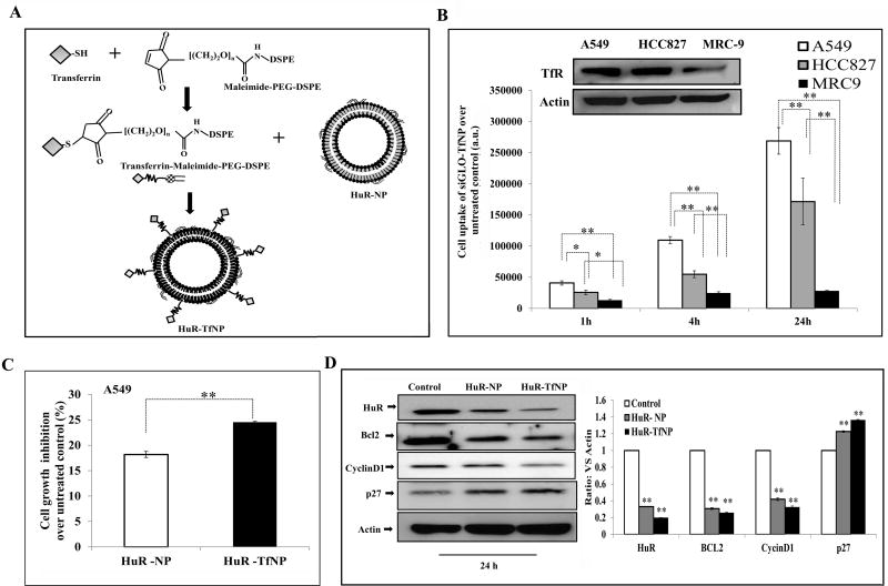
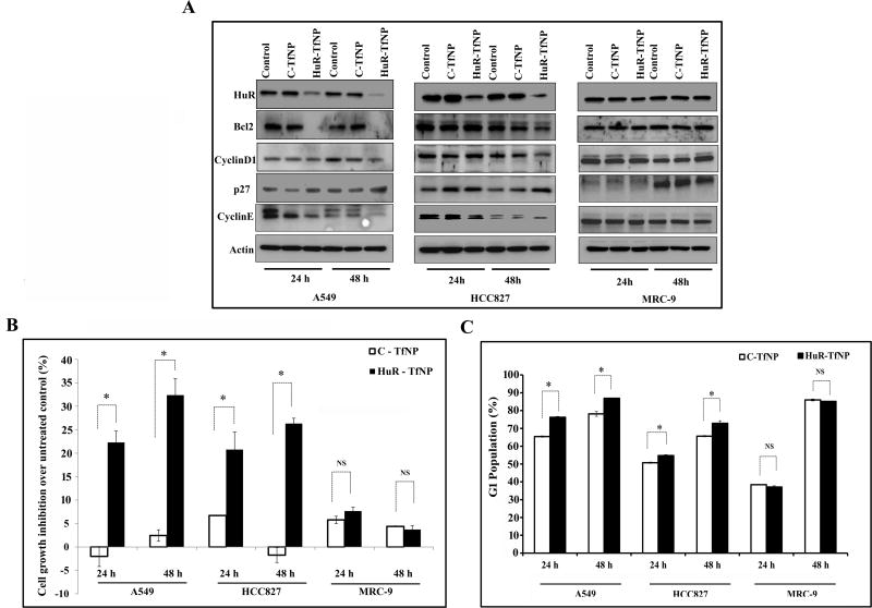
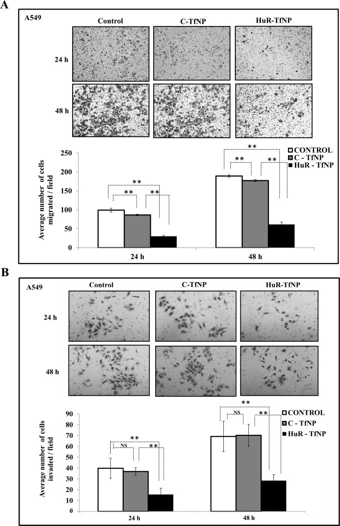
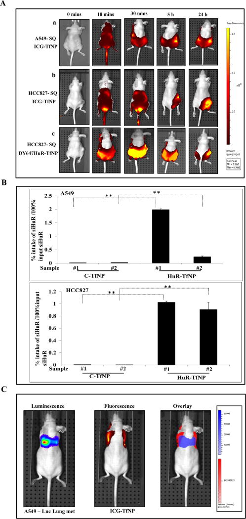
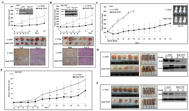
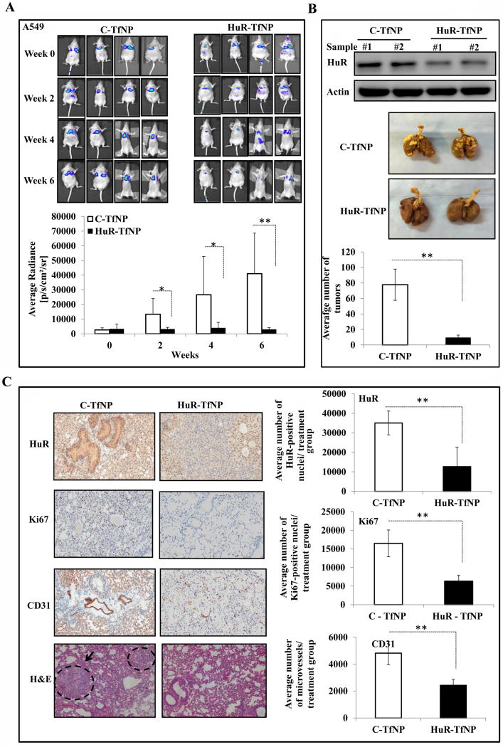
Selective downregulation of the human antigen R (HuR) protein by siRNA may provide a powerful approach for treating lung cancer. To this end, we investigated the efficacy of transferrin receptor-targeted liposomal nanoparticle-based HuR siRNA (HuR-TfNP) therapy and compared with control siRNA (C)-TfNP therapy both, in vitro and in vivo using lung cancer models. In vitro studies showed HuR-TfNP, but not C-TfNP, efficiently downregulated HuR and HuR-regulated proteins in A549, and HCC827 lung cancer cells, resulting in reduced cell viability, inhibition of cell migration and invasion, and induction of G1 cell-cycle arrest culminating in apoptosis. However, HuR-TfNP activity in normal MRC-9 lung fibroblasts was negligible. In vivo biodistribution study demonstrated that fluorescently labeled HuR-siRNA or ICG dye-loaded TfNP localized in tumor tissues. Efficacy studies showed intratumoral or intravenous administration of HuR-TfNP significantly inhibited A549 (>55% inhibition) and HCC827 (>45% inhibition) subcutaneous tumor growth compared with C-TfNP. Furthermore, HuR-TfNP treatment reduced HuR, Ki67, and CD31 expression and increased caspase-9 and PARP cleavage and TUNEL-positive staining indicative of apoptotic cell death in tumor tissues compared with C-TfNP treatment. The antitumor activity of HuR-TfNP was also observed in an A549-luc lung metastatic model, as significantly fewer tumor nodules (9.5 ± 3.1; P < 0.001; 88% inhibition) were observed in HuR-TfNP-treated group compared with the C-TfNP-treated group (77.7 ± 20.1). Significant reduction in HuR, Ki67, and CD31 expression was also observed in the tumor tissues of HuR-TfNP-treatment compared with C-TfNP treatment. Our findings highlight HuR-TfNP as a promising nanotherapeutic system for lung cancer treatment. Mol Cancer Ther; 16(8); 1470-86. ©2017 AACR.
©2017 American Association for Cancer Research.
Figures
Similar articles
-
Folate receptor-targeted nanoparticle delivery of HuR-RNAi suppresses lung cancer cell proliferation and migration.J Nanobiotechnology. 2016 Jun 21;14(1):47. doi: 10.1186/s12951-016-0201-1. J Nanobiotechnology. 2016. PMID: 27328938 Free PMC article.
-
HuR-targeted small molecule inhibitor exhibits cytotoxicity towards human lung cancer cells.Sci Rep. 2017 Aug 30;7(1):9694. doi: 10.1038/s41598-017-07787-4. Sci Rep. 2017. PMID: 28855578 Free PMC article.
-
HuR-targeted nanotherapy in combination with AMD3100 suppresses CXCR4 expression, cell growth, migration and invasion in lung cancer.Cancer Gene Ther. 2015 Dec;22(12):581-90. doi: 10.1038/cgt.2015.55. Epub 2015 Oct 23. Cancer Gene Ther. 2015. PMID: 26494555 Free PMC article.
-
Drug delivery approaches for HuR-targeted therapy for lung cancer.Adv Drug Deliv Rev. 2022 Jan;180:114068. doi: 10.1016/j.addr.2021.114068. Epub 2021 Nov 22. Adv Drug Deliv Rev. 2022. PMID: 34822926 Free PMC article. Review.
-
HuR-targeted agents: An insight into medicinal chemistry, biophysical, computational studies and pharmacological effects on cancer models.Adv Drug Deliv Rev. 2022 Feb;181:114088. doi: 10.1016/j.addr.2021.114088. Epub 2021 Dec 20. Adv Drug Deliv Rev. 2022. PMID: 34942276 Review.
Cited by
-
The RNA-binding protein HuR in human cancer: A friend or foe?Adv Drug Deliv Rev. 2022 May;184:114179. doi: 10.1016/j.addr.2022.114179. Epub 2022 Mar 3. Adv Drug Deliv Rev. 2022. PMID: 35248670 Free PMC article. Review.
-
Surmounting Cancer Drug Resistance: New Perspective on RNA-Binding Proteins.Pharmaceuticals (Basel). 2023 Aug 7;16(8):1114. doi: 10.3390/ph16081114. Pharmaceuticals (Basel). 2023. PMID: 37631029 Free PMC article. Review.
-
Enhanced Anti-tumor of Pep-1 Modified Superparamagnetic Iron Oxide/PTX Loaded Polymer Nanoparticles.Front Pharmacol. 2019 Jan 22;9:1556. doi: 10.3389/fphar.2018.01556. eCollection 2018. Front Pharmacol. 2019. PMID: 30723412 Free PMC article.
-
Molecular entrapment by RNA: an emerging tool for disrupting protein-RNA interactions in vivo.RNA Biol. 2020 Apr;17(4):417-424. doi: 10.1080/15476286.2020.1717059. Epub 2020 Jan 28. RNA Biol. 2020. PMID: 31957541 Free PMC article.
-
Therapeutic approaches targeting molecular signaling pathways common to diabetes, lung diseases and cancer.Adv Drug Deliv Rev. 2021 Nov;178:113918. doi: 10.1016/j.addr.2021.113918. Epub 2021 Aug 8. Adv Drug Deliv Rev. 2021. PMID: 34375681 Free PMC article. Review.
References
-
- Allen TM. Ligand-targeted therapeutics in anticancer therapy. Nat Rev Cancer. 2002;2:750–763. - PubMed
-
- Boult J, Roberts K, Brookes MJ, Hughes S, Bury JP, Cross SS, et al. Overexpression of cellular iron import proteins is associated with malignant progression of esophageal adenocarcinoma. Clin Cancer Res. 2008;14:379–387. - PubMed
-
- Whitney JF, Clark JM, Griffin TW, Gautam S, Leslie KO. Transferrin receptor expression in non-small cell lung cancer Histopathologic and clinical correlates. Cancer. 1995;76:20–25. - PubMed
Publication types
MeSH terms
Substances
Grants and funding
LinkOut - more resources
Full Text Sources
Other Literature Sources
Medical
Miscellaneous
