Editorial
. 2017 Jun 1;23(11):2617-2629.
doi: 10.1158/1078-0432.CCR-16-2810.

Mutational Signatures in Breast Cancer: The Problem at the DNA Level

Affiliations
Editorial

Mutational Signatures in Breast Cancer: The Problem at the DNA Level

Serena Nik-Zainal et al. Clin Cancer Res. .

Abstract

A breast cancer genome is a record of the historic mutagenic activity that has occurred throughout the development of the tumor. Indeed, every mutation may be informative. Although driver mutations were the main focus of cancer research for a long time, passenger mutational signatures, the imprints of DNA damage and DNA repair processes that have been operative during tumorigenesis, are also biologically illuminating. This review is a chronicle of how the concept of mutational signatures arose and brings the reader up-to-date on this field, particularly in breast cancer. Mutational signatures have now been advanced to include mutational processes that involve rearrangements, and novel cancer biological insights have been gained through studying these in great detail. Furthermore, there are efforts to take this field into the clinical sphere. If validated, mutational signatures could thus form an additional weapon in the arsenal of cancer precision diagnostics and therapeutic stratification in the modern war against cancer. Clin Cancer Res; 23(11); 2617-29. ©2017 AACRSee all articles in this CCR Focus section, "Breast Cancer Research: From Base Pairs to Populations."


Figures

Figure 1
Figure 1. Somatic mutational processes in human cancer.
Each mutational process leaves a characteristic imprint – or mutational signature – on the cancer genome, comprising DNA damage and DNA repair components. The arrows indicate the duration and intensity of exposure to a specific mutational process. The amount of exposure to each mutational process could vary from one person to another. Mutational processes A, B, C and D represent hypothetical mutational processes that have occurred through the lifetime of the developing tumour. A could represent a normal mutational process that happens in all our cells (including normal cells), hence it is occurring in a small amount throughout life. B could represent a mutational process caused by an environmental insult, such as an occupational exposure to a carcinogen. C could represent a mutational process which occurs in bursts through tumorigenesis such as intermitted exposure to a chemical or to an intermittent disease process. D could represent the acquisition of a defect in a gene involved in normal DNA repair. The final mutational portrait is a composite of all the mutational processes that have been active over the lifetime of the cancer patient. A different patient could have all of these mutational processes occurring in their tumour or could have some of the same mutational processes as well as other mutational processes present.
Figure 2
Figure 2. Currently known extracted substitution mutational signatures in human breast cancers
(A) Table of twelve mutation signatures extracted using Non-Negative Matrix Factorization. Each signature is ordered by mutation class (C>A/G>T, C>G/G>C, C>T/G>A, T>A/A>T, T>C/A>G, T>G/A>C), taking immediate flanking sequence into account resulting in 96 triplets. For each class, mutations are ordered by 5’ base (A, C, G, T) first before 3’ base (A, C, G, T). Y axis reports the probability of a signature generating each of the 96 triplets. Signature extraction was performed separately in seventeen cancer types. The bars report the results of the extraction on the 560 breast cancers [37] using a widely-available algorithm using simply default parameters (39), and the error bars demonstrate the variability (of the presumptive same signatures) between cancers of different tissue types. The table also contains the associated etiologies of each signature, the prevalence of these signatures in breast cancer and whether the signature is also seen in other tumor types. (B) Top panel shows absolute numbers of mutations of each signature in each sample. Lower panel shows proportion of each signature in each sample. Panel B reprinted by permission from Macmillan Publishers Ltd.: Nature 534:47–54, copyright 2016.
Figure 3
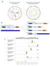
Figure 3. Extracting rearrangement mutational signatures in human breast cancers
(A) Whole genome circos plots were adapted from the R Circos package. Features depicted in circos plots from outermost rings heading inwards: Karyotypic ideogram outermost. Base substitutions next, plotted as rainfall plots (log10 intermutation distance on radial axis, dot colours: blue=C>A, black=C>G, red=C>T, grey=T>A, green=T>C, pink=T>G). Ring with short green lines = insertions, ring with short red lines = deletions. Major copy number allele (green = gain) ring, minor copy number allele ring (pink=loss), Central lines represent rearrangements (green= tandem duplications, pink=deletions, blue=inversions and gray=interchromosomal events. Note the difference in the nature of the distribution of rearrangements between the two tumors depicted. The whole genome profile on the left has >300 rearrangements which are clustered at distinct loci in specific chromosomes. By contrast, the >300 rearrangements present in the profile on the right-hand-side are uniformly dispersed through the genome. The mutational processes underpinning the differing distributions in these two tumors are most likely to be different. Thus, separating rearrangements into whether they are clustered or dispersed represents a first step in the rearrangement classification. (B) Types of rearrangements that can be ascertained easily. The hypothetical pieces of reference DNA from two different chromosomes on the left can be rearranged to form four main classes of rearrangements as shown on the right. This is a second step in the classification of rearrangements prior to rearrangement signature extraction. The rearrangements are also divided by size before extraction. (C) Six rearrangement signatures extracted using Non-Negative Matrix Factorization. Probability of rearrangement element on y-axis. Rearrangement size on x-axis. Del= deletion, tds = tandem duplication, inv = inversion, trans = translocation.
Figure 4
Figure 4. The spectrum of signatures within 560 breast cancers and individual patient whole genome profiles.
The panels in the middle represent from top to bottom: BRCA1 or BRCA2 null samples (dark purple) versus what is believed to be non-BRCA1/BRCA2 mutated samples (light purple), Estrogen Receptor (ER) status (black = positive, grey = negative), proportions of substitution signatures, rearrangement signatures and indel patterns present in the 560 patients. Figure legends are provided at the top of the figure. Samples are ordered according to hierarchical clustering performed on rearrangement mutational signatures. Six whole genome profiles of individual patients are shown to demonstrate how individualised each cancer genome is per patient. Note the striking differences between the six patients, even within the same “group” (Groups B and G). Group D is enriched with BRCA1-null tumors, Group G is enriched with BRCA2-null tumors and Group F is enriched with tumors that are never genetically BRCA1-null, are BRCA-like but different.
Figure 5
Figure 5. Mechanistic insights from mutagenesis: The APOBEC family of enzymes in genome-wide (Signatures 2 and 13) and localised mutational signatures (kataegis).
Based on the predominant cytosine mutagenesis at a TpC sequence context, the APOBEC family of enzymes has been implicated in causing both localised kataegis and genome-wide Signatures 2 and 13. (A) APOBECs cause DNA damage particularly on ssDNA, by deaminating cytosine into uracil. Uracil-N-glycosylase (UNG) first removes uracil before other components of the Base Excision Repair pathway restore the damaged DNA to its original state. If DNA is uncorrected and enters replication as uracil or an abasic site, then the possibilities are of generating C>T transition or C>G and C>A transversion mutations. (B) Although APOBECs are involved in both localised and genome-wide mutagenesis, there is mounting experimental and analytical evidence to support the hypotheses that these signatures arise by different mechanisms. Kataegis is believed to require a double-strand break (DSB) to arise first, before end-resection of the DSB leaves ssDNA exposed for APOBEC deamination (left-side panel). By contrast, APOBEC deamination that gives rise to Signatures 2 and 13 requires long stretches of ssDNA that could occur during uncoupling of the leading and lagging replication strands (right-side panel).

References

    1. Stratton MR, Campbell PJ, Futreal PA. The cancer genome. Nature. 2009;458(7239):719–24. - PMC - PubMed
    1. Stephens PJ, Tarpey PS, Davies H, Van Loo P, Greenman C, Wedge DC, et al. The landscape of cancer genes and mutational processes in breast cancer. Nature. 2012;486(7403):400–4. - PMC - PubMed
    1. Ching HC, Naidu R, Seong MK, Har YC, Taib NA. Integrated analysis of copy number and loss of heterozygosity in primary breast carcinomas using high-density SNP array. Int J Oncol. 2011;39(3):621–33. - PubMed
    1. Fang M, Toher J, Morgan M, Davison J, Tannenbaum S, Claffey K. Genomic differences between estrogen receptor (ER)-positive and ER-negative human breast carcinoma identified by single nucleotide polymorphism array comparative genome hybridization analysis. Cancer. 2011;117(10):2024–34. - PMC - PubMed
    1. Hicks J, Krasnitz A, Lakshmi B, Navin NE, Riggs M, Leibu E, et al. Novel patterns of genome rearrangement and their association with survival in breast cancer. Genome Res. 2006;16(12):1465–79. - PMC - PubMed

Publication types