. 2017 Sep;31(9):4129-4139.
doi: 10.1096/fj.201700140R. Epub 2017 Jun 1.

Complement-induced activation of MAPKs and Akt during sepsis: role in cardiac dysfunction

Affiliations

Complement-induced activation of MAPKs and Akt during sepsis: role in cardiac dysfunction

Fatemeh Fattahi et al. FASEB J. 2017 Sep.

Abstract

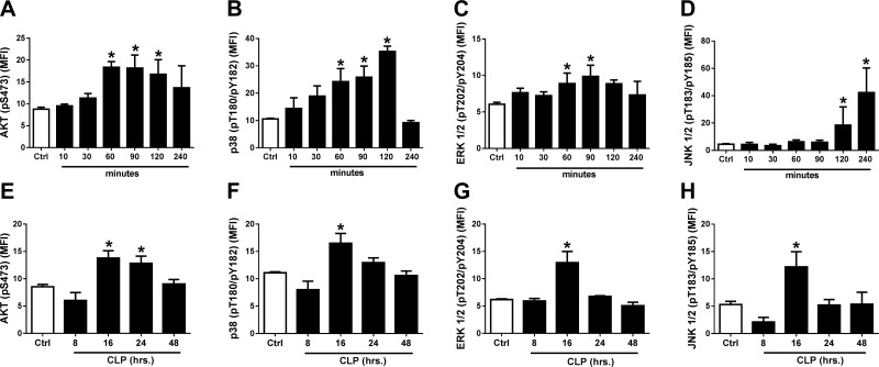
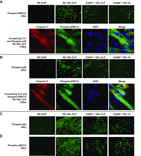
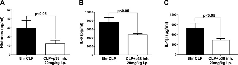
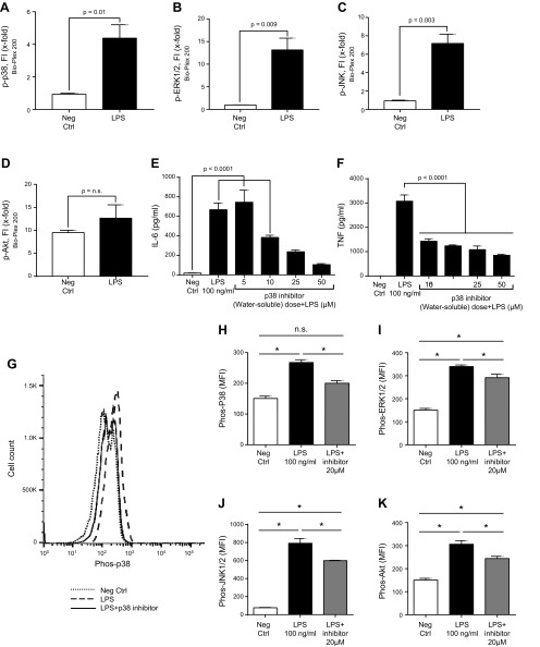
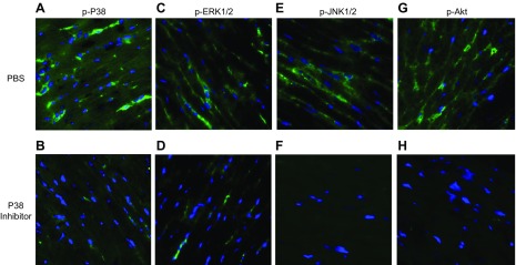
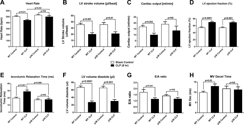
Polymicrobial sepsis in mice causes myocardial dysfunction after generation of the complement anaphylatoxin, complement component 5a (C5a). C5a interacts with its receptors on cardiomyocytes (CMs), resulting in redox imbalance and cardiac dysfunction that can be functionally measured and quantitated using Doppler echocardiography. In this report we have evaluated activation of MAPKs and Akt in CMs exposed to C5a in vitro and after cecal ligation and puncture (CLP) in vivo In both cases, C5a in vitro caused activation (phosphorylation) of MAPKs and Akt in CMs, which required availability of both C5a receptors. Using immunofluorescence technology, activation of MAPKs and Akt occurred in left ventricular (LV) CMs, requiring both C5a receptors, C5aR1 and -2. Use of a water-soluble p38 inhibitor curtailed activation in vivo of MAPKs and Akt in LV CMs as well as the appearance of cytokines and histones in plasma from CLP mice. When mouse macrophages were exposed in vitro to LPS, activation of MAPKs and Akt also occurred. The copresence of the p38 inhibitor blocked these activation responses. Finally, the presence of the p38 inhibitor in CLP mice reduced the development of cardiac dysfunction. These data suggest that polymicrobial sepsis causes cardiac dysfunction that appears to be linked to activation of MAPKs and Akt in heart.-Fattahi, F., Kalbitz, M., Malan, E. A., Abe, E., Jajou, L., Huber-Lang, M. S., Bosmann, M., Russell, M. W., Zetoune, F. S., Ward, P. A. Complement-induced activation of MAPKs and Akt during sepsis: role in cardiac dysfunction.

Keywords: C5a receptor; CLP; cardiomyocyte.


Figures

Figure 1.
Figure 1.
Time courses for C5a-induced (1 µg rrC5a/ml) in vitro activation (phosphorylation) of Akt (A, E) and the 3 MAPKs (p38, B, F; ERK-1/2, C, G; and JNK-1/2, D, H) in rat CMs. Using antibodies to phosphoamino acids in Akt or in MAPKs, end points were determined by flow cytometry in saponin-permeabilized rat CMs. AD) On the vertical axes are the antibody targets (phosphoserine, phosphothreonine, or phosphotyrosine). On the horizontal axes are the time courses (min) of activation. E–H) The time courses (h) for CLP-induced activation (phosphorylation) of Akt and MAPKs in permeabilized rat CMs as a function of time after CLP. MFI, mean fluorescence intensity, indicating levels of phospho-MAPK and phospho-Akt. Ctrl, control CMs from sham-treated rats. Data are expressed as means ± sem (n ≥ 6 for all time groups). *P < 0.05.
Figure 2.
Figure 2.
Phospho-MAPKs and Akt before (Ctrl, control) and 16 h after CLP, in frozen LV sections of WT mouse hearts or C5aR-KO mouse hearts. A) Phospho-ERK-1/2 before (Ctrl) or 16 h after CLP. Red: TnT; green: phospho-ERK-1/2; blue: DAPI-stained nuclei; yellow-green: merged image of TnT and ERK-1/2 labeling in CMs. A) Top: phospo-ERK-1/2 revealed green staining of CMs 16 h after CLP. The staining was markedly reduced in CLP CMs from C5aR-KO mice. B) Phospho-p38 images were very similar to those for phospho-ERK-1/2. C, D) Phospho-Akt (C) and phospho-JNK-1/2 (D) 16 h after CLP in WT CMs and CMs with and without C5aR1 and -R2. There were marked reductions in phospho-Akt and phospho-JNK-1/2 in frozen LV sections from C5aR-KO mice (n ≥ 3 for each group of frozen sections).
Figure 3.
Figure 3.
Treatment of CLP mice with a water-soluble p38 inhibitor, which was given 2 h before CLP. As measured by ELISA, there were sharp reductions 8 h after CLP in plasma biomarkers of sepsis: extracellular histones (A), IL-6 (B), and IL-1β (C) (n = 10 mice per marker). Data are expressed as means ± sem.
Figure 4.
Figure 4.
Phosphorylation of mouse PEMs (1 h at 37°C) caused by LPS (100 ng/ml). A–D) Bio-Plex phosphoprotein assay analysis of intracellular phospho-MAPKs p-p38 (A), p-ERK1/2 (B), and p-JNK (C) or p-AKT (D). Neg Ctrl, negative control. E, F) Dose response test of water-soluble p38 inhibitor (5–50 µM), in which the IC50 was used, indicated a dose-related reduction in release of IL-6 (E) and TNF (F). G–K) In vitro reductions in phospho-MAPKs and Akt in PEMs exposed to LPS in the absence or presence of 20 µM water-soluble inhibitor of p38. G) Typical example of PEMs exposed to buffer (neg ctrl) or to LPS in the presence or absence of the p38 inhibitor, using flow cytometry as the end point for phospho-Akt or phospho-MAPKs, as described in Fig. 1. HK) Levels of mean fluorescence intensity (MFI) indicating the levels of phospho-MAPKs p-p38 (H), p-ERK1/2 (I), and p-JNK (J) or p-AKT (K) in PEMs exposed to LPS in the absence or presence of 20 µM water-soluble inhibitor of p38. Data are expressed as means ± sem (n ≥ 5 samples per group). *P < 0.05.
Figure 5.
Figure 5.
Blockage of activation of MAPKs by water-soluble p38 inhibitor 16 h after CLP, determined with techniques similar to those described in Fig. 2. A, C, E, G) At 16 h after CLP, CMs demonstrated phosphorylation of MAPKs p38 (A), ERK1/2 (C), and p-JNK (E) and AKT (G). Green: MAPK; blue: DAPI. B, D, F, H) Pre-CLP administration of the p38 inhibitor blocked activation in CMs of all 3 MAPKs (p38, B; ERK1/2, D; and p-JNK, F) and AKT (H). (n = 4 separate samples in each group).
Figure 6.
Figure 6.
Echo Doppler parameters in hearts of mice before (WT control) and 8 h after CLP. Heart rate (A), LV stroke volume (B), cardiac output (C), LV ejection fraction (D), IVRT (E), LV volume diastole (F), E/A ratio (G), and MV deceleration time (H). Data are expressed as means ± sem (n ≥ 6 mice per group).

References

    1. Romero-Bermejo F. J., Ruiz-Bailen M., Gil-Cebrian J., Huertos-Ranchal M. J. (2011) Sepsis-induced cardiomyopathy. Curr. Cardiol. Rev. 7, 163–183 - PMC - PubMed
    1. Fernandes C. J. Jr., Akamine N., Knobel E. (1999) Cardiac troponin: a new serum marker of myocardial injury in sepsis. Intensive Care Med. 25, 1165–1168 - PubMed
    1. Blanco J., Muriel-Bombín A., Sagredo V., Taboada F., Gandía F., Tamayo L., Collado J., García-Labattut A., Carriedo D., Valledor M., De Frutos M., López M. J., Caballero A., Guerra J., Alvarez B., Mayo A., Villar J.; Grupo de Estudios y Análisis en Cuidados Intensivos (2008) Incidence, organ dysfunction and mortality in severe sepsis: a Spanish multicentre study. Crit. Care 12, R158 - PMC - PubMed
    1. Ward P. A., Guo R. F., Riedemann N. C. (2012) Manipulation of the complement system for benefit in sepsis. Crit. Care Res. Pract. 2012, 427607. - PMC - PubMed
    1. Nakae H., Endo S., Inada K., Takakuwa T., Kasai T., Yoshida M. (1994) Serum complement levels and severity of sepsis. Res. Commun. Chem. Pathol. Pharmacol. 84, 189–195 - PubMed

Publication types

MeSH terms