. 2017 Jul 7;357(6346):83-88.
doi: 10.1126/science.aam9243. Epub 2017 Jun 1.

Neurodevelopmental protein Musashi-1 interacts with the Zika genome and promotes viral replication

Affiliations

Neurodevelopmental protein Musashi-1 interacts with the Zika genome and promotes viral replication

Pavithra L Chavali et al. Science. .

Abstract

A recent outbreak of Zika virus in Brazil has led to a simultaneous increase in reports of neonatal microcephaly. Zika targets cerebral neural precursors, a cell population essential for cortical development, but the cause of this neurotropism remains obscure. Here we report that the neural RNA-binding protein Musashi-1 (MSI1) interacts with the Zika genome and enables viral replication. Zika infection disrupts the binding of MSI1 to its endogenous targets, thereby deregulating expression of factors implicated in neural stem cell function. We further show that MSI1 is highly expressed in neural progenitors of the human embryonic brain and is mutated in individuals with autosomal recessive primary microcephaly. Selective MSI1 expression in neural precursors could therefore explain the exceptional vulnerability of these cells to Zika infection.


Figures

Fig. 1
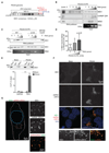
Fig. 1. MSI1 interacts directly with the ZIKV RNA genome.
A) Schematic diagram of PE243 ZIKV containing three putative MSI1 binding sites in its 3’UTR. Sites shared with MR766 are red, site unique to PE243 is blue. B) RNA pull-down assays performed with the 3’UTR of PE243. Increasing concentrations of in vitro transcribed biotinylated PE243 RNA were incubated with U-251 cell extracts and RNA-protein complexes were captured on streptavidin beads. Representative western blots were probed with antibodies against MSI1 and the RNA binding proteins, Musashi-2 (MSI2) and hnRNP Q/R. Corresponding protein and RNA inputs are shown on right. C) RNA pull-down assays performed with the wild type (WT) or triple mutant (Δ123) 3’UTR of PE243. Note that PE243-3’UTR_Δ123 lacks all three MSI1 binding sites depicted in Fig. 1A (see Fig. S1C for further details). Increasing concentrations of in vitro transcribed biotinylated RNA were incubated with U-251 cell extracts and RNA-protein complexes were captured on streptavidin beads. Representative western blots probed with antibody against MSI1 are shown together with corresponding protein and RNA inputs. D) Densitometric analysis of MSI1 levels from western blots of RNA pull-down assays, an example for which is shown in Fig. 1C. The amount of MSI1 precipitated by PE243-3’UTR_Δ123 is expressed as a percentage of MSI1 precipitated by the same concentration of PE243-3’UTR_WT. n=3 biological replicates. P-values were obtained from Student’s t-test, unpaired, two-tailed: * p<0.05, *** p<0.0005. Bar charts depict mean±s.e.m. E) CLIP analysis from mock- or ZIKV PE243-infected U-251 cells. Western blot shows immunoprecipitatations (IP) by rabbit IgG and MSI1 antibodies from mock and PE243-infected U-251 cells following UV crosslinking. Input (5%) represents whole cell extract. Western blot was probed with MSI1 antibody. Graph below shows qPCR performed on bound RNA from IP. CLIP values are presented as a percentage of input following subtraction of the IgG background. GAPDH serves as negative control. n=3 biological replicates. Note that a primer pair against 9519-9681 bp of ZIKV genome was used in these qPCRs (Table S4). F) Immunofluorescence of mock- or PE243-infected U-251 cells. MSI1 (green) and dsRNA (red) signals are detected by confocal microscopy. DNA is detected by Hoechst stain (blue). Framed area is shown at higher magnification below. G) Immunofluorescence of a PE243-infected U-251 cell. MSI1 (green) and dsRNA (red) signals are detected by STED super resolution microscopy. Outlines of the cell and nucleus are indicated in white and blue, respectively. Framed area is shown at higher magnification.
Fig. 2
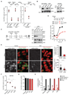
Fig. 2. MSI1 is required for effective replication of ZIKV.
A) Graph on left depicts viral RNA copies in control siRNA- (siCon) and MSI1 siRNA (siMSI1)-treated SK-N-BE2c and U-251 cells following infection with PE243 (MOI: 1 FFU/cell, 72h). Graph on right shows viral RNA copies in MSI1 siRNA-treated H9-derived neural stem cells (NSC) following infection with PE243 (MOI: 1 FFU/cell, 48h). Note that in all viral replication assays ZIKV was quantified by TaqMan assay as described in Materials and Methods. n=3 biological replicates. P-values were obtained from Student’s t-test, unpaired, two-tailed: * p<0.05, ** p<0.005, not significant (n.s.). B) Representative western blots of cell lines treated with control and MSI1 siRNAs from Fig. 2A. Blots were probed with antibodies against MSI1 or p150 as loading control. C) Location of the guide RNAs used for CRISPR/Cas9-mediated editing of the MSI1 locus in U-251 cells. For further details see text and Fig. S2. Western blots of parental, control, KO1 and KO2 cell lines probed with antibodies against N- and C-termini of MSI1 (NT or CT) and MSI2. p150 serves as loading control. D) Kinetics of PE243 viral RNA copies following infection in U-251 cells of different genotypes at the indicated time points. Note that cell death precluded collection of RNA from parental and control cells at 72h (MOI: 3 FFU/cell, 72h). E) Confocal microscopy images of PE243-infected control and KO1 U-251 cells immunostained with antibodies against dsRNA (green) and Hoechst DNA stain (red) following mock or PE243 infection (MOI: 3 FFU/cell, 48h). Graph on top shows percentage of cells containing dsRNA signal, whereas box plot on bottom depicts total dsRNA staining volume per cell. Note that only cells with detectable dsRNA signal were included in the latter analysis. Boxes: 25th to 75th percentile; whiskers: 5-95% range; line: median. P-value represents Student’s t-test, unpaired, two-tailed: **** p<0.0001. F) Viral RNA copies in U-251 cells of different genotypes following infection with MR766 (MOI: 3 FFU/cell, 48 h). n=3 biological replicates. P-values were obtained from Student’s t-test, unpaired, two-tailed: * p<0.05, ** p<0.005, not significant (n.s.). G) Virus-binding assays performed under conditions that prevent internalisation. PE243 infection was performed in U-251 cells of different genotypes (MOI: 3 FFU/cell, 1h). n=3 biological replicates. P-values were obtained from Student’s t-test, unpaired, two-tailed: * p<0.05, ** p<0.005, not significant (n.s.). Bar charts depict mean±s.e.m. H) Pseudotyped particle infectivity assay in U-251 cells of different genotypes. HIV (pNL4-3.luc.R-E-) or MoMLV pseudotyped virus expressing a luciferase reporter, with either PE243 ZIKV, VSVg or a negative control envelope used to determine viral entry events. n=3 biological replicates. P-values were obtained from Student’s t-test, unpaired, two-tailed: * p<0.05, ** p<0.005, not significant (n.s.). Bar charts depict mean±s.e.m.
Fig. 3
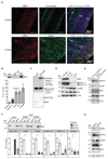
Fig. 3. MSI1 is enriched in neural progenitors, regulates Microcephalin (MCPH1) expression and is mutated in MCPH patients.
A) Immunohistochemistry of human embryonic brain at post conception week (pcw) 10 and 12. Tissue sections stained with antibodies against MSI1 (red) combined with neuron-specific β-III tubulin (green), or the apical neural progenitor marker Nestin (green). DNA is detected by DAPI (blue). Note that MSI1 is enriched in neural progenitors at the ventricular and subventricular zones (VZ and SVZ), but is absent from the cortical plate (CP). B) MSI1 CLIP from U-251 cells with genotypes as indicated. CLIP was performed with rabbit IgG or MSI1 antibodies. Graphs show qPCRs of bound transcripts. n=3 biological replicates. P-values were obtained from Student’s t-test, unpaired, two-tailed: * p<0.05, ** p<0.005, not significant (n.s). C) RNA EMSA analysis to detect binding between MCPH1_L 3’UTR and purified GST-MSI1 recombinant protein. Coomassie staining of corresponding purified proteins is shown below. D) Western blots of parental, control, KO1 and KO2 cell lines. Blots were probed with antibodies as indicated. p150 serves as loading control. E) Western blots of whole cell lysates from parent-of-patient- and patient-derived primary lymphocytes. Blots were probed with antibodies as indicated. MSI1 levels are unchanged. F) MSI1 RIP from mock- and ZIKV-infected U-251 cells (MOI: 1 FFU/cell). Rabbit IgG or MSI1 antibodies were used for immunoprecipitation. Input corresponds to 10% of whole cell extract. Western blot was probed with MSI1 antibody. Graphs below show amounts of bound RNAs including ZIKV genome and endogenous target transcripts. RIP values are presented as a percentage of input following subtraction of the IgG background. MR766 and PE243 were quantitated by TaqMan assay, whereas endogenous transcripts with SYBR qPCR. Bar charts depict mean±s.e.m. n=3 biological replicates. P-values were obtained from Student’s t-test, unpaired, two-tailed: * p<0.05, ** p<0.005, not significant (n.s.). G) Western blots of PE243-infected U-251 cells. (MOI: 1 FFU/cell, 72h). Blots were probed with antibodies as indicated. α-tubulin serves as loading control. Note that MSI1 positively regulates MCPH1 and CDK6, and negatively regulates NUMB and p21 protein levels.
Fig. 4
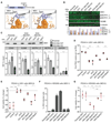
Fig. 4. The A184V MCPH mutation disrupts RNA binding by MSI1 and impairs the ability of MSI1 to drive ZIKV replication.
A) Structural model of MSI1 (blue) and HRP1/RNA complex (grey/orange) predicts that the A184V mutation impairs the interaction with RNA due to a steric clash. B) Western blots of cell lines stably expressing WT or A184V MSI1 transgenes. Note that cell lines were derived from either KO1 or KO2 cells as specified. Blots were probed with antibodies as indicated. p150 serves as loading control. Graph shows signal intensities of each protein normalized to parental cells. C) MSI1 RIP from U-251 cells with genotypes as indicated. Rabbit IgG or MSI1 antibodies were used for immunoprecipitation. Input corresponds to 10% of whole cell extract. Western blot was probed with MSI1 antibody. Graphs below show qPCRs of bound transcripts. n=3 biological replicates. P-values were obtained from Student’s t-test, unpaired, two-tailed: * p<0.05, ** p<0.005, not significant (n.s). D) Quantification of viral RNA copies in U-251 cells with the indicated genotypes following infection with PE243 (MOI: 3 FFU/cell, 48h). n=3 biological replicates. P-values were obtained from Student’s t-test, unpaired, two-tailed: ** p<0.005, not significant (n.s). E) Changes in survival of U-251 cells with different genotypes following infection with PE243 (MOI: 3 FFU/cell, 48h). n=3 biological replicates. P-values were obtained from Student’s t-test, unpaired, two-tailed: * p<0.05, ** p<0.005, not significant (n.s). F) ZIKV infection of HEK293T cells as measured by FACS analysis of flavivirus protein E. FACS was performed on control, MsiWT or MsiAV transgene-expressing cells following infection with PE243 (MOI: 1 FFU/cell, 48h). n=2 biological replicates. Bar charts depict mean±s.e.m. G) Changes in survival of control, MsiWT or MsiAV transgene-expressing HEK293T cells following infection with PE243 (MOI: 3 FFU/cell, 48h). n=3 biological replicates, and P-values were obtained from Student’s t-test, unpaired, two-tailed: * p<0.05, ** p<0.005, not significant (n.s).

Comment in

Similar articles

Cited by

References

    1. Mlakar J, et al. Zika Virus Associated with Microcephaly. The New England journal of medicine. 2016;374:951–958. - PubMed
    1. Oliveira Melo AS, et al. Zika virus intrauterine infection causes fetal brain abnormality and microcephaly: tip of the iceberg? Ultrasound in obstetrics & gynecology : the official journal of the International Society of Ultrasound in Obstetrics and Gynecology. 2016;47:6–7. - PubMed
    1. Cugola FR, et al. The Brazilian Zika virus strain causes birth defects in experimental models. Nature. 2016;534:267–271. - PMC - PubMed
    1. Slavov SN, Otaguiri KK, Kashima S, Covas DT. Overview of Zika virus (ZIKV) infection in regards to the Brazilian epidemic. Brazilian journal of medical and biological research = Revista brasileira de pesquisas medicas e biologicas / Sociedade Brasileira de Biofisica … [et al.] 2016;49:e5420. - PMC - PubMed
    1. Cauchemez S, et al. Association between Zika virus and microcephaly in French Polynesia, 2013-15: a retrospective study. Lancet. 2016;387:2125–2132. - PMC - PubMed

Publication types

MeSH terms