

Clinical or ATPase domain mutations in ABCD4 disrupt the interaction between the vitamin B12-trafficking proteins ABCD4 and LMBD1
- PMID: 28572511
- PMCID: PMC5512089
- DOI: 10.1074/jbc.M117.784819
Clinical or ATPase domain mutations in ABCD4 disrupt the interaction between the vitamin B12-trafficking proteins ABCD4 and LMBD1
Abstract
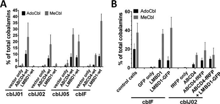
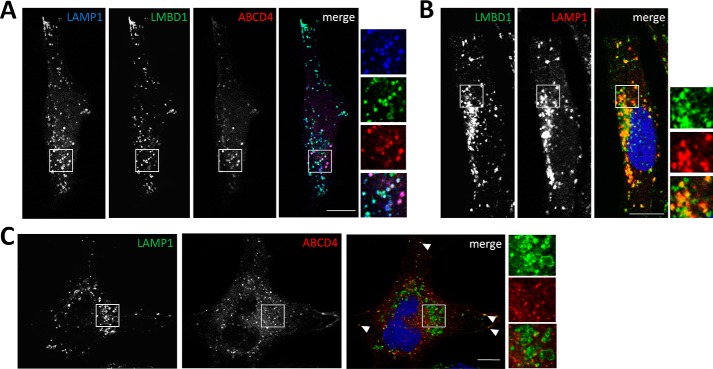
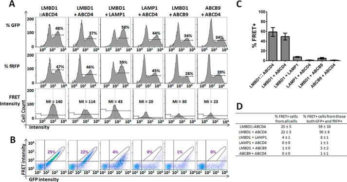
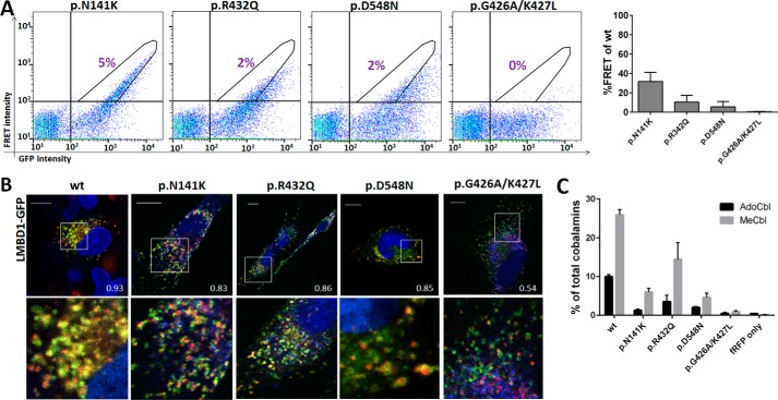
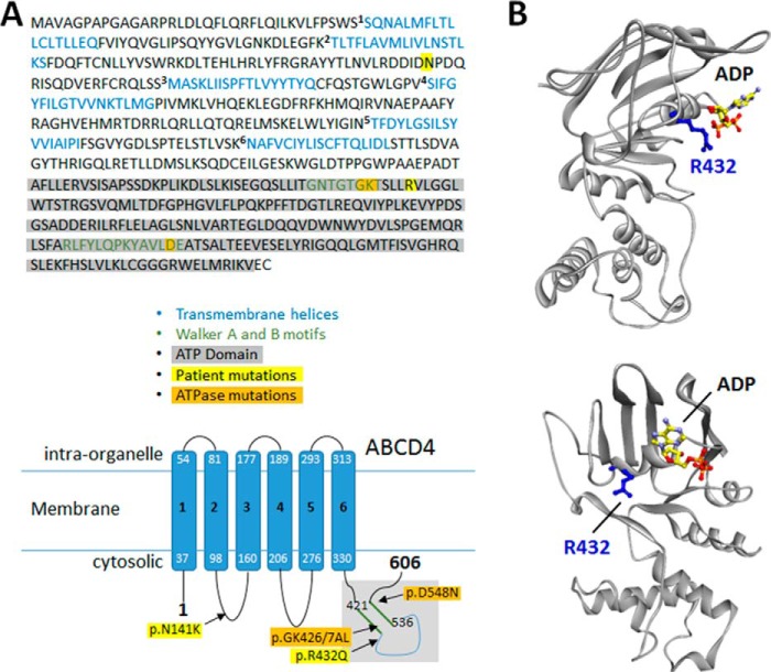
Vitamin B12 (cobalamin (Cbl)), in the cofactor forms methyl-Cbl and adenosyl-Cbl, is required for the function of the essential enzymes methionine synthase and methylmalonyl-CoA mutase, respectively. Cbl enters mammalian cells by receptor-mediated endocytosis of protein-bound Cbl followed by lysosomal export of free Cbl to the cytosol and further processing to these cofactor forms. The integral membrane proteins LMBD1 and ABCD4 are required for lysosomal release of Cbl, and mutations in the genes LMBRD1 and ABCD4 result in the cobalamin metabolism disorders cblF and cblJ. We report a new (fifth) patient with the cblJ disorder who presented at 7 days of age with poor feeding, hypotonia, methylmalonic aciduria, and elevated plasma homocysteine and harbored the mutations c.1667_1668delAG [p.Glu556Glyfs*27] and c.1295G>A [p.Arg432Gln] in the ABCD4 gene. Cbl cofactor forms are decreased in fibroblasts from this patient but could be rescued by overexpression of either ABCD4 or, unexpectedly, LMBD1. Using a sensitive live-cell FRET assay, we demonstrated selective interaction between ABCD4 and LMBD1 and decreased interaction when ABCD4 harbored the patient mutations p.Arg432Gln or p.Asn141Lys or when artificial mutations disrupted the ATPase domain. Finally, we showed that ABCD4 lysosomal targeting depends on co-expression of, and interaction with, LMBD1. These data broaden the patient and mutation spectrum of cblJ deficiency, establish a sensitive live-cell assay to detect the LMBD1-ABCD4 interaction, and confirm the importance of this interaction for proper intracellular targeting of ABCD4 and cobalamin cofactor synthesis.
Keywords: ABC transporter; ABCD4; LMBD1; cblF; cblJ; fluorescence resonance energy transfer (FRET); homology modeling; inborn error of metabolism; protein-protein interaction; vitamin B12.
© 2017 by The American Society for Biochemistry and Molecular Biology, Inc.
Conflict of interest statement
The authors declare that they have no conflicts of interest with the contents of this article
Figures
Similar articles
-
Purification and interaction analyses of two human lysosomal vitamin B12 transporters: LMBD1 and ABCD4.Mol Membr Biol. 2014 Nov-Dec;31(7-8):250-61. doi: 10.3109/09687688.2014.990998. Mol Membr Biol. 2014. PMID: 25535791
-
The lysosomal protein ABCD4 can transport vitamin B12 across liposomal membranes in vitro.J Biol Chem. 2021 Jan-Jun;296:100654. doi: 10.1016/j.jbc.2021.100654. Epub 2021 May 3. J Biol Chem. 2021. PMID: 33845046 Free PMC article.
-
Mutations in ABCD4 cause a new inborn error of vitamin B12 metabolism.Nat Genet. 2012 Oct;44(10):1152-5. doi: 10.1038/ng.2386. Epub 2012 Aug 26. Nat Genet. 2012. PMID: 22922874
-
Insights into lysosomal cobalamin trafficking: lessons learned from cblF disease.J Mol Med (Berl). 2010 May;88(5):459-66. doi: 10.1007/s00109-010-0601-x. Epub 2010 Feb 20. J Mol Med (Berl). 2010. PMID: 20174775 Review.
-
Genetic defects of folate and cobalamin metabolism.Eur J Pediatr. 1998 Apr;157 Suppl 2:S60-6. doi: 10.1007/pl00014306. Eur J Pediatr. 1998. PMID: 9587028 Review.
Cited by
-
Inherited disorders of lysosomal membrane transporters.Biochim Biophys Acta Biomembr. 2020 Dec 1;1862(12):183336. doi: 10.1016/j.bbamem.2020.183336. Epub 2020 May 8. Biochim Biophys Acta Biomembr. 2020. PMID: 32389669 Free PMC article. Review.
-
A QTL for Number of Teats Shows Breed Specific Effects on Number of Vertebrae in Pigs: Bridging the Gap Between Molecular and Quantitative Genetics.Front Genet. 2019 Mar 26;10:272. doi: 10.3389/fgene.2019.00272. eCollection 2019. Front Genet. 2019. PMID: 30972109 Free PMC article.
-
The role of vitamin B12 in viral infections: a comprehensive review of its relationship with the muscle-gut-brain axis and implications for SARS-CoV-2 infection.Nutr Rev. 2022 Feb 10;80(3):561-578. doi: 10.1093/nutrit/nuab092. Nutr Rev. 2022. PMID: 34791425 Free PMC article. Review.
-
Redox-Linked Coordination Chemistry Directs Vitamin B12 Trafficking.Acc Chem Res. 2021 Apr 20;54(8):2003-2013. doi: 10.1021/acs.accounts.1c00083. Epub 2021 Apr 2. Acc Chem Res. 2021. PMID: 33797888 Free PMC article.
-
Assessment of cellular cobalamin metabolism in Gaucher disease.BMC Med Genet. 2020 Jan 13;21(1):12. doi: 10.1186/s12881-020-0947-z. BMC Med Genet. 2020. PMID: 31931749 Free PMC article.
References
-
- Coelho D., Kim J. C., Miousse I. R., Fung S., du Moulin M., Buers I., Suormala T., Burda P., Frapolli M., Stucki M., Nürnberg P., Thiele H., Robenek H., Höhne W., Longo N., Pasquali M., et al. (2012) Mutations in ABCD4 cause a new inborn error of vitamin B12 metabolism. Nat. Genet. 44, 1152–1155 - PubMed
-
- Rutsch F., Gailus S., Miousse I. R., Suormala T., Sagné C., Toliat M. R., Nürnberg G., Wittkampf T., Buers I., Sharifi A., Stucki M., Becker C., Baumgartner M., Robenek H., Marquardt T., et al. (2009) Identification of a putative lysosomal cobalamin exporter altered in the cblF defect of vitamin B12 metabolism. Nat. Genet. 41, 234–239 - PubMed
Publication types
MeSH terms
Substances
Supplementary concepts
Associated data
- Actions
- Actions
LinkOut - more resources
Full Text Sources
Other Literature Sources
Medical
Molecular Biology Databases
Research Materials
Miscellaneous
