Comparative Study
. 2017 Jul 21;292(29):12089-12099.
doi: 10.1074/jbc.M117.787622. Epub 2017 Jun 1.

Domain alternation and active site remodeling are conserved structural features of ubiquitin E1

Affiliations
Comparative Study

Domain alternation and active site remodeling are conserved structural features of ubiquitin E1

Zongyang Lv et al. J Biol Chem. .

Abstract

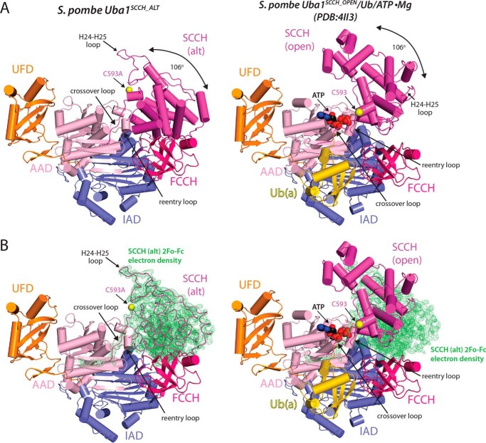
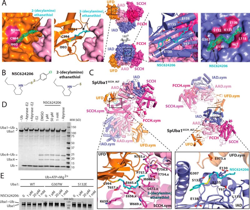
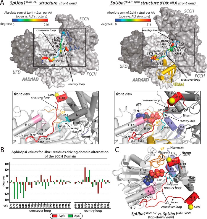
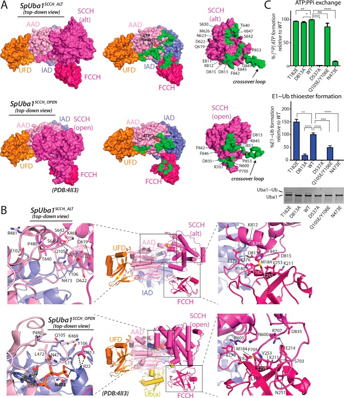
E1 enzymes for ubiquitin (Ub) and Ub-like modifiers (Ubls) harbor two catalytic activities that are required for Ub/Ubl activation: adenylation and thioester bond formation. Structural studies of the E1 for the Ubl small ubiquitin-like modifier (SUMO) revealed a single active site that is transformed by a conformational switch that toggles its competency for catalysis of these two distinct chemical reactions. Although the mechanisms of adenylation and thioester bond formation revealed by SUMO E1 structures are thought to be conserved in Ub E1, there is currently a lack of structural data supporting this hypothesis. Here, we present a structure of Schizosaccharomyces pombe Uba1 in which the second catalytic cysteine half-domain (SCCH domain) harboring the catalytic cysteine has undergone a 106° rotation that results in a completely different network of intramolecular interactions between the SCCH and adenylation domains and translocation of the catalytic cysteine 12 Å closer to the Ub C terminus compared with previous Uba1 structures. SCCH domain alternation is accompanied by conformational changes within the Uba1 adenylation domains that effectively disassemble the adenylation active site. Importantly, the structural and biochemical data suggest that domain alternation and remodeling of the adenylation active site are interconnected and are intrinsic structural features of Uba1 and that the overall structural basis for adenylation and thioester bond formation exhibited by SUMO E1 is indeed conserved in Ub E1. Finally, the mechanistic insights provided by the novel conformational snapshot of Uba1 presented in this study may guide efforts to develop small molecule inhibitors of this critically important enzyme that is an active target for anticancer therapeutics.

Keywords: E1; X-ray crystallography; adenylation; conformational change; domain alternation; enzyme mechanism; enzyme structure; structure-function; thioester; ubiquitin.


Conflict of interest statement

The authors declare that they have no conflicts of interest with the contents of this article

Figures

Figure 1.
Figure 1.
Crystal structure of domain-alternated S. pombe Uba1. A, comparison of the Uba1SCCH_ALT (left) and Uba1SCCH_OPEN (Protein Data Bank code 4II3, right) structures, shown as ribbon diagrams. Uba1 domains are labeled and color-coded, and ATP and Ub(a) from the Uba1SCCH_OPEN structure are shown as spheres and a yellow ribbon diagram, respectively. Catalytic cysteines positions (Cys593) are indicated with an arrow, and the SCCH domain alternation is highlighted by a double-headed arrow. B, ribbon diagrams of the Uba1SCCH_ALT (left) and Uba1SCCH_OPEN (right) structures overlaid with the 2FoFc electron density (green) for the SCCH domain of the Uba1SCCH_ALT structure. For clarity, the SCCH domain of the Uba1SCCH_ALT structure is shown as loops. The Uba1 domains, ATP, and Ub(a) are colored and labeled as in A. alt, domain-alternated SCCH conformation.
Figure 2.
Figure 2.
Interactions between the Uba1SCCH_ALT and NSC624206. A, middle, the Uba1SCCH_ALT structure is shown as a surface representation in two orientations, and the NSC624206 compound noncovalently bound to the IAD and the 2-(decylamino)ethanethiol fragment covalently bound to the UFD via disulfide bond are shown as sticks. The domains are colored and labeled as in Fig. 1. Left, magnified view of the 2-(decylamino)ethanethiol fragment of NSC624206, shown as sticks, bound to the UFD. Omit 2FoFc electron density for the fragment is shown in green. The UFD and AAD are shown as surface representations, and residues that form contacts with the fragment are colored and labeled. Second from left, magnified view of the 2-(decylamino)ethanethiol fragment, shown as sticks, bound to Cys994 of the UFD, shown as a ribbon diagram. Side chains of residues that form contacts with the fragment are shown as sticks and labeled. The AAD is shown as a surface representation. Second from right, magnified view of the NSC624206 compound, shown as sticks, bound to the IAD, shown as a ribbon diagram. Side chains of residues that form contacts with the compound are shown as sticks and labeled. The IAD and SCCH are shown as surface representations. Right, magnified view of the NSC624206 compound, shown as sticks, bound to the IAD. Omit 2FoFc electron density for the compound is shown in green. The IAD is shown as a surface representation, and residues that form contacts with the fragment are colored and labeled. B, chemical structures of the NSC624206 compound and the 2-(decylamino)ethanethiol fragment. C, crystal contacts with the NSC624206 compound. Top, the Uba1SCCH_ALT structure is shown as a ribbon diagram in two different orientations, and the domains are colored and labeled as in A. Symmetry (sym) mates that form contacts with the 2-(decylamino)ethanethiol fragment, shown as sticks, bound to the UFD (left) and to the NSC624206 compound, shown as sticks, bound to the IAD (right) are also shown as semitransparent ribbon diagrams with the domains colored and labeled. Bottom, magnified views of the 2-(decylamino)ethanethiol fragment bound to the UFD (left) and NSC624206 compound bound to the IAD (right). The side chains of residues from Uba1SCCH_ALT and the symmetry mates that form contacts with the fragment or compound are shown as sticks and labeled. The .x indicates residues residing in symmetry-related Uba1 molecules. D, biochemical analysis of the effect of 2-(decylamino)ethanethiol modification on UFD Cys994. An E1-E2 single turnover thioester transfer assay from Uba1 to Ubc4 was performed with different concentrations of NSC624206 or DMSO (see “Experimental procedures” for details). E, biochemical analysis of the inhibitory effect of NSC624206 on WT and mutant Uba1 activity. Uba1∼Ub thioester formation assays were performed as readouts of Uba1 catalytic activity with different concentrations of NSC624206 or DMSO (see “Experimental procedures” for details).
Figure 3.
Figure 3.
Active site remodeling in S. pombe Uba1. A, conformational changes in the crossover and reentry loops upon SCCH domain alternation. Top, Uba1 from the Uba1SCCH_ALT and Uba1SCCH_open structures is shown as a surface representation with indicated domains colored various shades of gray. Ub(a) from the Uba1SCCH_OPEN structure is shown as a gold ribbon diagram. The crossover and reentry loops are shown as putty representations, and the diameter and color (see color code above structures) of the putty correspond to the sum of the absolute change in φ and ψ angles. Bottom, active site remodeling accompanies Uba1 SCCH domain alternation. The Uba1SCCH_ALT (left) and Uba1SCCH_OPEN (right) structures are shown in the same orientation as ribbon diagrams with domains colored as in the top panels. Regions undergoing conformational changes in the Uba1 active site upon SCCH domain alternation are color-coded and labeled. H1 and H2 of Uba1 that become disordered in the Uba1SCCH_ALT structure are shown as semitransparent slate spheres. The α-carbons of the residues that undergo significant changes between the open and closed conformations are labeled and shown as spheres. B, differences in φ and ψ angles for residues in the crossover and reentry loops between the Uba1SCCH_ALT and Uba1SCCH_OPEN structures were calculated and plotted. C, the adenylation domains of the Uba1SCCH_ALT and Uba1SCCH_OPEN structures were superimposed and are shown as ribbon diagrams. ATP from the Uba1SCCH_OPEN structure is shown as spheres. Note that this panel is presented in a top-down view, which differs from the front views presented in A to better highlight the conformational changes that occur during active site remodeling and SCCH domain alternation in the context of the superimposed structures. AA, amino acid; alt, domain-alternated SCCH conformation.
Figure 4.
Figure 4.
Comparison of Uba1 and SUMO E1 in different conformational states. A, the Uba1SCCH_OPEN (left) and Uba1SCCH_ALT (right) structures are shown as surface representations (with the exception of the SCCH domain, which is shown as a light magenta ribbon diagram) with domains color-coded and labeled. Ub(a) and ATP were modeled onto the Uba1SCCH_ALT structure based on how they interact with Uba1 in the Uba1SCCH_OPEN structure. SCCH domain helices are labeled, and the N- and C-terminal ends of the helices are indicated to facilitate structure comparison. H18 and H19 of the Uba1 structures are colored red to correspond with H6 and H7 of the SUMO E1 structures in B. H29 of the Uba1 structures is colored cyan to correspond with H13 of the SUMO E1 structures in B. The approximate direction of the SCCH domain alternation is indicated with a double-headed arrow. A magnified view of the Uba1SCCH_ALT active site is shown in the right inset. B, the SUMO E1SCCH_OPEN (Protein Data Bank code 3KYC) and SUMO E1SCCH_CLOSED (Protein Data Bank code 3KYD) structures are presented in the same style and orientation as Uba1 in A. C, comparison of the crossover/reentry loops and SCCH domains in the Uba1SCCH_OPEN (Protein Data Bank code 4II3) and SUMO E1SCCH_OPEN (Protein Data Bank code 3KYC) structures (bottom) and in the Uba1SCCH_ALT and SUMO E1SCCH_CLOSED (Protein Data Bank code 3KYD) structures (top). The adenylation domains of the structures were superimposed. The structures are shown as ribbon diagrams and ATP, 5′-(vinylsulfonylaminodeoxy)adenosine (AVSN), or 5′-(sulfamoylaminodeoxy)adenosine (AMSN) are shown as sticks. The difference in SCCH domain positioning in the Uba1SCCH_ALT and SUMO E1SCCH_CLOSED structures is indicated with a double-headed arrow. D, left, comparison of the Uba1 and SUMO E1 SCCH domains. Unique structural elements of the Uba1 SCCH domain are indicated with a black dashed oval. Right, the Uba1 SCCH domain was docked onto Uba1 in the same orientation as the SUMO E1SCCH_CLOSED structure. Steric clashes involving the unique structural elements of the Uba1 SCCH domain illustrate the fact that the extent of domain alternation in Uba1 and SUMO E1 during thioester bond formation is unlikely to be precisely the same. alt, domain-alternated SCCH conformation; SUMO(a), adenylated SUMO.
Figure 5.
Figure 5.
A unique network of contacts among the Uba1 SCCH, FCCH, and AAD domains in the Uba1SCCH_ALT structure. A, Uba1SCCH_ALT (top left) and Uba1SCCH_OPEN (bottom left) structures are shown as surface representations with domains labeled and color-coded. For clarity, Ub(a) from the Uba1SCCH_OPEN structure is not shown. The right panels show the intramolecular interface between Uba1 and the globular SCCH domain as an open book representation with residues buried at the interface shaded green. To highlight the altered network of interactions resulting from the SCCH domain alternation, SCCH residues involved in unique intramolecular interactions in the two structures are labeled. B, Uba1SCCH_ALT (top) and Uba1SCCH_OPEN (bottom) structures are shown as ribbon diagrams with magnified views of the SCCH/AAD and SCCH/FCCH intramolecular interfaces shown in the left and right insets, respectively. Residues involved in interactions are shown as sticks, and hydrogen bonds are indicated with dashed black lines. C, structure-function analysis of intramolecular contacts between Uba1 and the globular SCCH domain unique to the Uba1SCCH_ALT structure. ATP/PPi exchange assays (top) and Uba1∼Ub thioester formation assays (bottom) were performed as readouts of Uba1 catalytic activities. All experiments were performed in triplicate as described under “Experimental procedures,” and error bars represent ±1 S.D. *, p ≤ 0.05; **, p < 0.01; ***, p < 0.001; ****, p < 0.0001; ns, not significant. alt, domain-alternated SCCH conformation.

References

    1. Komander D., and Rape M. (2012) The ubiquitin code. Annu. Rev. Biochem. 81, 203–229 - PubMed
    1. van der Veen A. G., and Ploegh H. L. (2012) Ubiquitin-like proteins. Annu. Rev. Biochem. 81, 323–357 - PubMed
    1. Hochstrasser M. (2009) Origin and function of ubiquitin-like proteins. Nature 458, 422–429 - PMC - PubMed
    1. Haas A. L., and Rose I. A. (1982) The mechanism of ubiquitin activating enzyme. A kinetic and equilibrium analysis. J. Biol. Chem. 257, 10329–10337 - PubMed
    1. Haas A. L., Warms J. V., Hershko A., and Rose I. A. (1982) Ubiquitin-activating enzyme. Mechanism and role in protein-ubiquitin conjugation. J. Biol. Chem. 257, 2543–2548 - PubMed

Publication types

MeSH terms

LinkOut - more resources