

Photoacoustic imaging of tumor targeting with riboflavin-functionalized theranostic nanocarriers
- PMID: 28572726
- PMCID: PMC5441666
- DOI: 10.2147/IJN.S125192
Photoacoustic imaging of tumor targeting with riboflavin-functionalized theranostic nanocarriers
Abstract
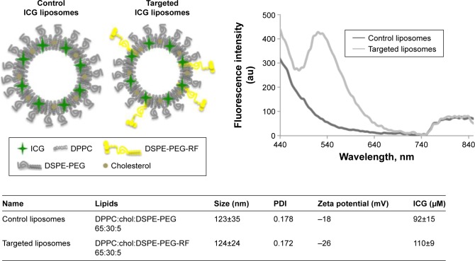
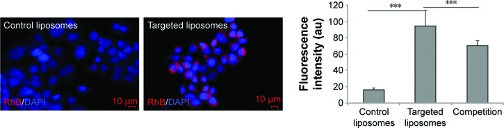
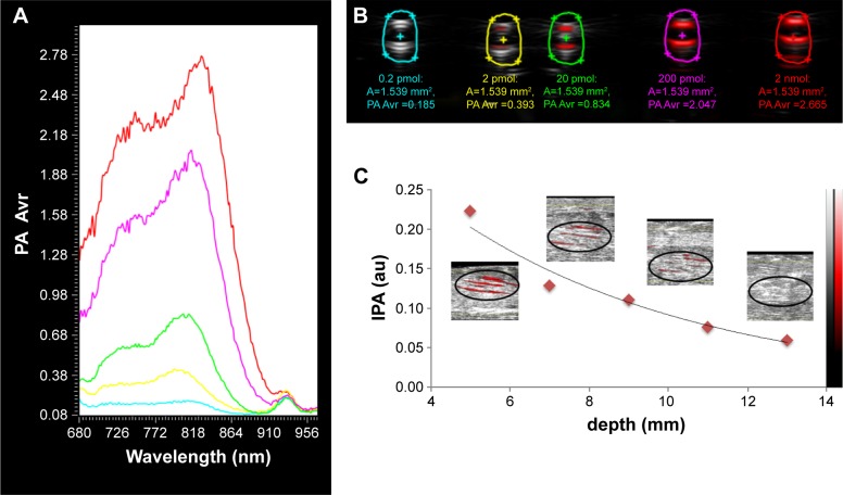
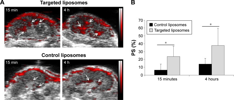
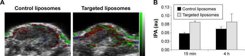
Photoacoustic imaging is an emerging method in the molecular imaging field, providing high spatiotemporal resolution and sufficient imaging depths for many clinical applications. Therefore, the aim of this study was to use photoacoustic imaging as a tool to evaluate a riboflavin (RF)-based targeted nanoplatform. RF is internalized by the cells through a specific pathway, and its derivatives were recently shown as promising tumor-targeting vectors for the drug delivery systems. Here, the RF amphiphile synthesized from a PEGylated phospholipid was successfully inserted into a long-circulating liposome formulation labeled with the clinically approved photoacoustic contrast agent - indocyanine green (ICG). The obtained liposomes had a diameter of 124 nm (polydispersity index =0.17) and had a negative zeta potential of -26 mV. Studies in biological phantoms indicated a stable and concentration-dependent photoacoustic signal (Vevo® LAZR) of the ICG-containing RF-functionalized liposomes. In A431 cells, a high uptake of RF-functionalized liposomes was found and could be blocked competitively. First, studies in mice revealed ~3 times higher photoacoustic signal in subcutaneous A431 tumor xenografts (P<0.05) after injection of RF-functionalized liposomes compared to control particles. In this context, the application of a spectral unmixing protocol confirmed the initial quantitative data and improved the localization of liposomes in the tumor. In conclusion, the synthesized RF amphiphile leads to efficient liposomal tumor targeting and can be favorably detected by photoacoustic imaging with a perspective of theranostic applications.
Keywords: active targeting; indocyanine green; long-circulating liposomes; photoacoustic imaging; riboflavin.
Conflict of interest statement
Disclosure The authors report no conflicts of interest in this work.
Figures
References
-
- Wang L, Yang PP, Zhao XX, Wang H. Self-assembled nanomaterials for photoacoustic imaging. Nanoscale. 2016;8:2488–2509. - PubMed
-
- James ML, Gambhir SS. A molecular imaging primer: modalities, imaging agents, and applications. Physiol Rev. 2012;92:897–965. - PubMed
-
- Beziere N, Lozano N, Nunes A, et al. Dynamic imaging of PEGylated indocyanine green (ICG) liposomes within the tumor microenvironment using multi-spectral optoacoustic tomography (MSOT) Biomaterials. 2015;37:415–424. - PubMed
-
- Luke GP, Yeager D, Emelianov SY. Biomedical applications of photoacoustic imaging with exogenous contrast agents. Ann Biomed Eng. 2012;40:422–437. - PubMed
MeSH terms
Substances
LinkOut - more resources
Full Text Sources
Other Literature Sources
