

Cytosolic and Nuclear Co-localization of Betalain Biosynthetic Enzymes in Tobacco Suggests that Betalains Are Synthesized in the Cytoplasm and/or Nucleus of Betalainic Plant Cells
- PMID: 28572813
- PMCID: PMC5435750
- DOI: 10.3389/fpls.2017.00831
Cytosolic and Nuclear Co-localization of Betalain Biosynthetic Enzymes in Tobacco Suggests that Betalains Are Synthesized in the Cytoplasm and/or Nucleus of Betalainic Plant Cells
Abstract
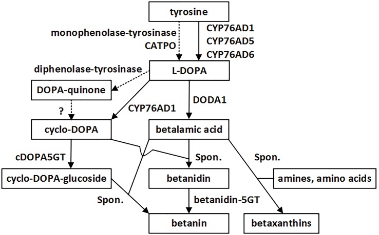
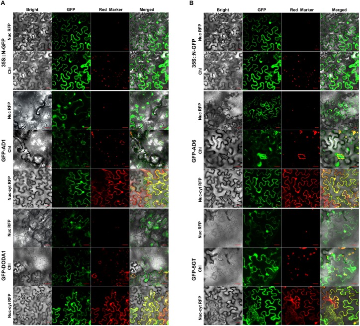
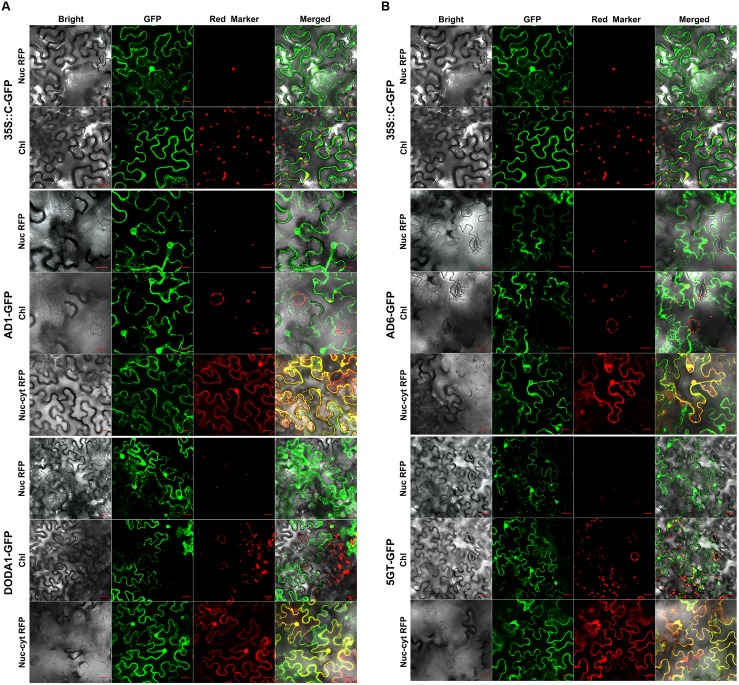
Betalains replace anthocyanins as color pigments in most families of Caryophyllales. Unlike anthocyanins, betalains are derived from tyrosine via three enzymatic steps: hydroxylation of L-tyrosine to L-3,4-dihydroxyphenylalanine (L-DOPA; step 1), and conversion of L-DOPA to betalamic acid (step 2), and to cyclo-DOPA (cDOPA; step 3). The principal enzymes responsible for these reactions have been elucidated at the molecular level, but their subcellular localizations have not been explored; hence, the intracellular compartments wherein betalains are biosynthesized remain unknown. Here, we report on the subcellular localization of these principal enzymes. Bioinformatic predictors and N- and C-terminal GFP tagging in transgenic tobacco, showed that Beta vulgaris CYP76AD1 which mediates both steps 1 and 3, DODA1 that catalyzes step 2, and CYP76AD6 which also mediates step 1, were similarly localized to the cytoplasm and nucleus (although the P450s were also weakly present in the endoplasmic reticulum). These two compartments were also the principal locations of Mirabilis jalapa cDOPA5GT. The cytoplasmic and nuclear co-localization of these key enzymes in tobacco suggests that betalains are biosynthesized in the cytoplasm and/or nucleus of betalain-containing plant cells. Elucidation of the subcellular compartmentation of betalain biosynthesis will facilitate the bioengineering of the betalain biosynthetic pathway in non-betalain-containing plants.
Keywords: CYP76AD1; DODA1 (DOD); betalain biosynthesis; cDOPA5GT; cytoplasm; nucleus; subcellular compartment; subcellular localization.
Figures
Similar articles
-
Elucidation of the first committed step in betalain biosynthesis enables the heterologous engineering of betalain pigments in plants.New Phytol. 2016 Apr;210(1):269-83. doi: 10.1111/nph.13796. Epub 2015 Dec 18. New Phytol. 2016. PMID: 26683006
-
Betalain production is possible in anthocyanin-producing plant species given the presence of DOPA-dioxygenase and L-DOPA.BMC Plant Biol. 2012 Mar 12;12:34. doi: 10.1186/1471-2229-12-34. BMC Plant Biol. 2012. PMID: 22409631 Free PMC article.
-
Gain-of-function mutations in beet DODA2 identify key residues for betalain pigment evolution.New Phytol. 2018 Jul;219(1):287-296. doi: 10.1111/nph.15159. Epub 2018 May 13. New Phytol. 2018. PMID: 29754447
-
The evolution of betalain biosynthesis in Caryophyllales.New Phytol. 2019 Oct;224(1):71-85. doi: 10.1111/nph.15980. Epub 2019 Jul 19. New Phytol. 2019. PMID: 31172524 Review.
-
Biotechnological production and emerging applications of betalains: A review.Biotechnol Adv. 2025 Jul-Aug;81:108576. doi: 10.1016/j.biotechadv.2025.108576. Epub 2025 Apr 9. Biotechnol Adv. 2025. PMID: 40204005 Review.
Cited by
-
Soybean Cyst Nematode-Resistant Protein AATRhg1 Affects Amino Acid Homeostasis and Betalain Accumulation.Plant Direct. 2025 Aug 26;9(8):e70098. doi: 10.1002/pld3.70098. eCollection 2025 Aug. Plant Direct. 2025. PMID: 40874116 Free PMC article.
-
Regulation Mechanism of Plant Pigments Biosynthesis: Anthocyanins, Carotenoids, and Betalains.Metabolites. 2022 Sep 16;12(9):871. doi: 10.3390/metabo12090871. Metabolites. 2022. PMID: 36144275 Free PMC article. Review.
-
A Genome-Wide Identification Study Reveals That HmoCYP76AD1, HmoDODAα1 and HmocDOPA5GT Involved in Betalain Biosynthesis in Hylocereus.Genes (Basel). 2021 Nov 23;12(12):1858. doi: 10.3390/genes12121858. Genes (Basel). 2021. PMID: 34946807 Free PMC article.
-
Optimizing Promoters and Subcellular Localization for Constitutive Transgene Expression in Marchantia polymorpha.Plant Cell Physiol. 2024 Sep 3;65(8):1298-1309. doi: 10.1093/pcp/pcae063. Plant Cell Physiol. 2024. PMID: 38822700 Free PMC article.
-
Enhanced thermotolerance of Arabidopsis by chitooligosaccharides-induced CERK1n-ERc fusion gene.Plant Signal Behav. 2020 Dec 1;15(12):1816322. doi: 10.1080/15592324.2020.1816322. Epub 2020 Sep 9. Plant Signal Behav. 2020. PMID: 32902365 Free PMC article.
References
LinkOut - more resources
Full Text Sources
Other Literature Sources