

No Functional Role for microRNA-342 in a Mouse Model of Pancreatic Acinar Carcinoma
- PMID: 28573106
- PMCID: PMC5435746
- DOI: 10.3389/fonc.2017.00101
No Functional Role for microRNA-342 in a Mouse Model of Pancreatic Acinar Carcinoma
Abstract
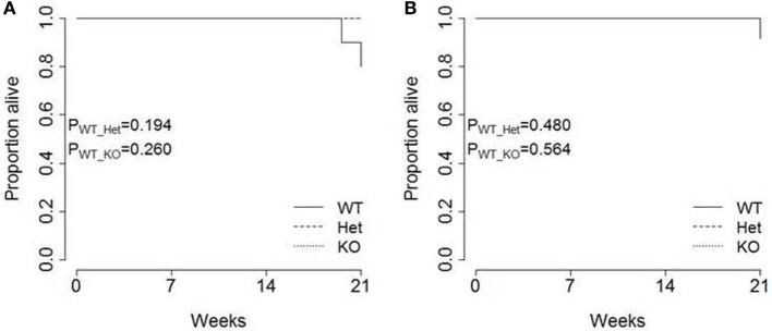
The intronic microRNA (miR)-342 has been proposed as a potent tumor-suppressor gene. miR-342 is found to be downregulated or epigenetically silenced in multiple different tumor sites, and this loss of expression permits the upregulation of several key oncogenic pathways. In several different cell lines, lower miR-342 expression results in enhanced proliferation and metastasis potential, both in vitro and in xenogenic transplant conditions. Here, we sought to determine the function of miR-342 in an in vivo spontaneous cancer model, using the Ela1-TAg transgenic model of pancreatic acinar carcinoma. Through longitudinal magnetic resonance imaging monitoring of Ela1-TAg transgenic mice, either wild-type or knockout for miR-342, we found no role for miR-342 in the development, growth rate, or pathogenicity of pancreatic acinar carcinoma. These results indicate the importance of assessing miR function in the complex physiology of in vivo model systems and indicate that further functional testing of miR-342 is required before concluding it is a bona fide tumor-suppressor-miR.
Keywords: acinar carcinoma; in vivo; miR-342-5p; microRNAs; pancreatic cancer.
Figures


Similar articles
-
miR-29a-deficiency does not modify the course of murine pancreatic acinar carcinoma.Oncotarget. 2017 Apr 18;8(16):26911-26917. doi: 10.18632/oncotarget.15850. Oncotarget. 2017. PMID: 28460473 Free PMC article.
-
Murine Pancreatic Acinar Cell Carcinoma Growth Kinetics Are Independent of Dietary Vitamin D Deficiency or Supplementation.Front Oncol. 2017 Jun 28;7:133. doi: 10.3389/fonc.2017.00133. eCollection 2017. Front Oncol. 2017. PMID: 28702373 Free PMC article.
-
MicroRNA-127 is aberrantly downregulated and acted as a functional tumor suppressor in human pancreatic cancer.Tumour Biol. 2016 Oct;37(10):14249-14257. doi: 10.1007/s13277-016-5270-0. Epub 2016 Aug 29. Tumour Biol. 2016. PMID: 27571739
-
MiRNA-615-5p functions as a tumor suppressor in pancreatic ductal adenocarcinoma by targeting AKT2.PLoS One. 2015 Apr 9;10(4):e0119783. doi: 10.1371/journal.pone.0119783. eCollection 2015. PLoS One. 2015. PMID: 25856297 Free PMC article.
-
MiR-132 promotes the proliferation, invasion and migration of human pancreatic carcinoma by inhibition of the tumor suppressor gene PTEN.Prog Biophys Mol Biol. 2019 Nov;148:65-72. doi: 10.1016/j.pbiomolbio.2017.09.019. Epub 2017 Sep 20. Prog Biophys Mol Biol. 2019. PMID: 28941804 Review.
Cited by
-
MiR-342 controls Mycobacterium tuberculosis susceptibility by modulating inflammation and cell death.EMBO Rep. 2021 Sep 6;22(9):e52252. doi: 10.15252/embr.202052252. Epub 2021 Jul 20. EMBO Rep. 2021. PMID: 34288348 Free PMC article.
-
microRNA-342-3p targets FOXQ1 to suppress the aggressive phenotype of nasopharyngeal carcinoma cells.BMC Cancer. 2019 Jan 24;19(1):104. doi: 10.1186/s12885-018-5225-5. BMC Cancer. 2019. PMID: 30678643 Free PMC article.
-
Aberrantly reduced expression of miR-342-5p contributes to CCND1-associated chronic myeloid leukemia progression and imatinib resistance.Cell Death Dis. 2021 Oct 5;12(10):908. doi: 10.1038/s41419-021-04209-2. Cell Death Dis. 2021. PMID: 34611140 Free PMC article.
-
Knockout of Acinar Enriched microRNAs in Mice Promote Duct Formation But Not Pancreatic Cancer.Sci Rep. 2019 Jul 31;9(1):11147. doi: 10.1038/s41598-019-47566-x. Sci Rep. 2019. PMID: 31367007 Free PMC article.
-
Serum miR-342-3p is a novel diagnostic and prognostic biomarker for non-small cell lung cancer.Int J Clin Exp Pathol. 2018 May 1;11(5):2742-2748. eCollection 2018. Int J Clin Exp Pathol. 2018. PMID: 31938391 Free PMC article.
References
LinkOut - more resources
Full Text Sources
Other Literature Sources