Review
. 2017:2017:7354260.
doi: 10.1155/2017/7354260. Epub 2017 May 10.

Colorectal Cancer: From the Genetic Model to Posttranscriptional Regulation by Noncoding RNAs

Affiliations
Review

Colorectal Cancer: From the Genetic Model to Posttranscriptional Regulation by Noncoding RNAs

María Antonia Lizarbe et al. Biomed Res Int. 2017.

Abstract

Colorectal cancer is the third most common form of cancer in developed countries and, despite the improvements achieved in its treatment options, remains as one of the main causes of cancer-related death. In this review, we first focus on colorectal carcinogenesis and on the genetic and epigenetic alterations involved. In addition, noncoding RNAs have been shown to be important regulators of gene expression. We present a general overview of what is known about these molecules and their role and dysregulation in cancer, with a special focus on the biogenesis, characteristics, and function of microRNAs. These molecules are important regulators of carcinogenesis, progression, invasion, angiogenesis, and metastases in cancer, including colorectal cancer. For this reason, miRNAs can be used as potential biomarkers for diagnosis, prognosis, and efficacy of chemotherapeutic treatments, or even as therapeutic agents, or as targets by themselves. Thus, this review highlights the importance of miRNAs in the development, progression, diagnosis, and therapy of colorectal cancer and summarizes current therapeutic approaches for the treatment of colorectal cancer.


Figures

Figure 1
Figure 1
Evolution of the number of publications on miRNAs and lncRNAs. (a) The number of publications per year (searched in PubMed) that contain the terms “microRNA” (black bars) and “microRNA + cancer” (empty bars) in their title, abstract, or keywords, is shown. The inset is equivalent with the terms “lncRNA” and “lncRNA + cancer”. (b) Evolution of the number of entries in miRBase from December 2002 until the last release (release 21) I June 2014. The inset shows the increase in the number of species where miRNAs have been found.
Figure 2
Figure 2
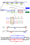
Interaction between miRNAs and target mRNAs in animals and miRNA-200 family. (a) miRNAs (green) target mRNAs (blue) in the 3′UTR. This mRNA region can bind one or various miRISC complexes arising from the same or different miRNAs. A perfect complementarity is found in the seed region between nucleotides 2 and 7 from the 5′ end of the miRNA (orange box). A central bulge prevents endonucleolytic cleavage mediated by Ago2 (a major difference with the miRNA-target mRNA interaction in plants). A few nucleotide matches in the miRNA 3′ end (especially between nucleotides 13 and 16; yellow box) are necessary for the best stabilization of the miRNA/mRNA duplex. The presence in the mRNA sequence of an A residue in position 1 and/or an A or U residue in position 9 can increase miRNA efficiency [6]. (b) The miR-200 family of miRNAs consists of two closely related subfamilies that differ in one nucleotide within the seed sequence (boxed). The five miR-200 family members are located on two different genetic loci in chromosomes 1 and 12. (c) Schematic representation of the secondary structure of the pre-miR-141 hairpin. The sequence of the mature miR-141-3p is indicated in red and the miR-141-5p (previously called miR-141) is in blue. Black triangles show Dicer cleavage sites due to its RNase III activity.
Figure 3
Figure 3
Biogenesis of miRNAs and translational repression exerted by miRNAs and siRNAs. The nascent pri-miRNA transcripts are first processed into ~70-nucleotide pre-miRNAs by Drosha/DGR8 complexes inside the nucleus. MiRNAs can also be byproducts of mRNA splicing after lariat debranching and 3′-trimming by the exosome complex PM/Scl (mirtrons). Pre-miRNAs (or mitrons) are transported to the cytoplasm by exportin 5 coupled with Ran-GTP and are processed into miRNA:miRNA duplexes by Dicer/TRBP. Dicer also processes endogenous or exogenous dsRNA duplexes. Only one strand of the miRNA:miRNA duplex or the siRNA duplex is preferentially assembled into RISC by the RISC loading complex (RLC). Subsequently, the RISC complex acts on its mRNA target by translational repression or mRNA cleavage, depending, at least in part, on the level of complementarity between the small RNA and its target. Alterations of miRNA function in cancer are multifactorial. They can arise from epigenetic silencing of miRNA genes or may be due to genetic instability as human microRNA genes are frequently located at fragile sites and genomic regions involved in cancers [7, 8]. Dysregulation of miRNA biogenesis machinery is also frequent in cancer mainly due to mutations in one or several of the proteins involved in processing (Drosha, DGCR8, DICER, TRBP, and Argonaute), or in nuclear export (exportin 5), or by alterations in their posttranslational modifications (PTMs) [, –14]. Although specific miRNAs have been described as acting as oncogenes and tumor suppressors, the miRNA expression profile of human tumors is characterized by a general defect in miRNA production that results in global miRNA downregulation. In addition, miRNA sequestration by the so-called miRNA sponges (i.e., circRNAs and lncRNAs) can also contribute to dysregulation of miRNA function. ORF, open reading frame.
Figure 4
Figure 4
Mechanisms of target regulation by miRNAs. miRNAs regulate gene expression through multiple pathways. Eukaryotic initiation factors bind the 5′cap and the cytoplasmic poly(A)-binding protein (PABPC), connecting the 5′ and 3′ ends of mRNAs and stimulating their translation by the ribosome. (a) The miRNA-induced silencing complex (miRISC) can induce translational repression by blocking initiation; GW182 competes with eIF-4G in association with PABPC; and Argonaute (AGO) binds to the mRNA cap releasing eIF-4E, thus preventing the circularization required for efficient translation and the binding of ribosome 40S subunit to the mRNA. (b) Translational repression can also be induced by the miRISC by inhibiting a step after initiation, such as promoting ribosome drop-off or stimulating proteolysis of the nascent peptide. (c) Partial pairing of the miRNA complex to target 3′UTR sites can result in deadenylation of the mRNA by the CCR4–NOT or the PAN2/3 complexes and decapping by DCP1/2 (all of them recruited by GW182). Loss of the poly(A) tail causes dissociation of PABPC and leads to degradation of the mRNA. (d) Finally, perfect pairing between a miRNA and its target site induces endonucleolytic cleavage by AGO, leading to rapid degradation of the mRNA (occurs mainly in plants) [15].
Figure 5
Figure 5
Chemical modifications of miRNA-targeting modulators. Anti-miR oligonucleotides have been chemically modified in order to achieve better stability against serum nucleases and to increase the binding affinity to targeted miRNAs. Most of the modifications are at the 2′ position of the sugar moiety, as 2′-O-methyl (2′-OMe), 2′-O-methoxyethyl (2′-MOE), and 2′-fluoro (2′-F). Locked nucleic acid (LNA) is a bicyclic RNA analogue in which ribose is locked by introduction of a methylene bridge between the 2′ oxygen and the 4′ carbon of the pentose. In addition, most anti-miR oligonucleotides contain phosphorothioate backbone linkages in which sulfur replaces one of the nonbridging oxygen atoms in the phosphate group. Morpholino oligomers replace the ribose with a methylenemorpholine ring (to which bases are attached) with phosphorodiamidate linkages. PNA oligomers are oligonucleotide analogues in which the ribose-phosphate backbone has been replaced with a peptide-like structure containing N-(2-aminoethyl)-glycine units. Both morpholino and PNA oligomers are uncharged, which facilitates the interaction with targeted miRNAs.

Similar articles

Cited by

References

    1. Arends M. J. Pathways of colorectal carcinogenesis. Applied Immunohistochemistry and Molecular Morphology. 2013;21(2):97–102. doi: 10.1097/PAI.0b013e3182849808. - DOI - PubMed
    1. Barrasa J. I., Olmo N., Lizarbe M. A., Turnay J. Bile acids in the colon, from healthy to cytotoxic molecules. Toxicology in Vitro. 2013;27(2):964–977. doi: 10.1016/j.tiv.2012.12.020. - DOI - PubMed
    1. Tariq K., Ghias K. Colorectal cancer carcinogenesis: a review of mechanisms. Cancer Biology and Medicine. 2016;13(1):120–135. doi: 10.28092/j.issn.2095-3941.2015.0103. - DOI - PMC - PubMed
    1. Tejpar S., Van Cutsem E. Molecular and genetic defects in colorectal tumorigenesis. Bailliere's Best Practice and Research in Clinical Gastroenterology. 2002;16(2):171–185. doi: 10.1053/bega.2001.0279. - DOI - PubMed
    1. Worthley D. L., Whitehall V. L., Spring K. J., Leggett B. A. Colorectal carcinogenesis: road maps to cancer. World Journal of Gastroenterology. 2007;13(28):3784–3791. doi: 10.3748/wjg.v13.i28.3784. - DOI - PMC - PubMed

MeSH terms

LinkOut - more resources