. 2017 Apr 25;3(5):e157.
doi: 10.1097/TXD.0000000000000666. eCollection 2017 May.

Modeling the Effect of the Aryl Hydrocarbon Receptor on Transplant Immunity

Affiliations

Modeling the Effect of the Aryl Hydrocarbon Receptor on Transplant Immunity

Walker Julliard et al. Transplant Direct. .

Abstract

Background: Exposure to pollutants through inhalation is a risk factor for lung diseases including cancer, asthma, and lung transplant rejection, but knowledge of the effects of inhaled pollutants on pathologies outside of the lung is limited.

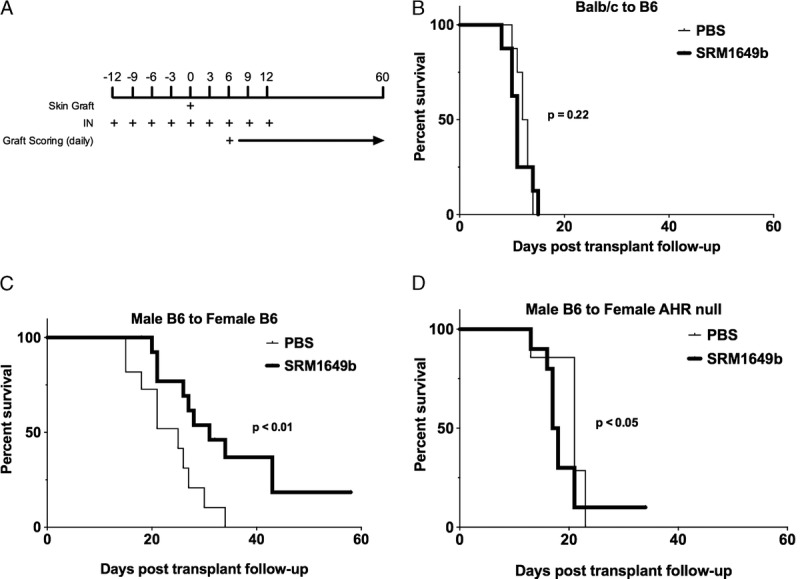
Methods: Using the minor-mismatched model of male C57BL/6J (B6) to female B6 skin grafts, recipient mice were treated with an inhaled urban dust particle sample every 3 days before and after grafting. Graft survival time was determined, and analysis of the resulting immune response was performed at time before rejection.

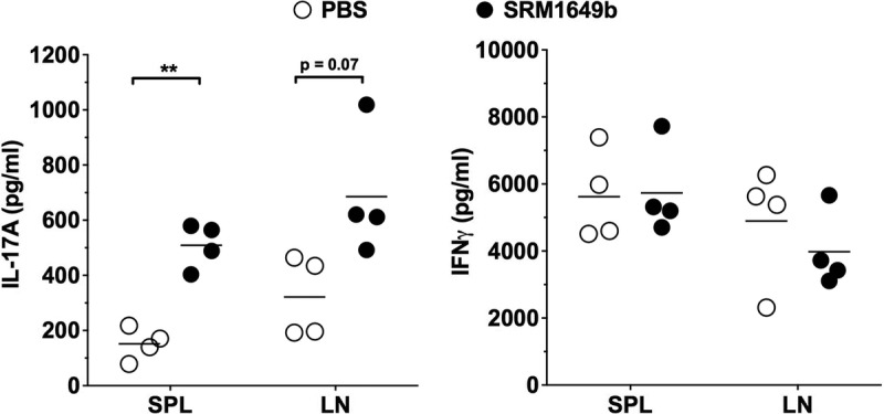
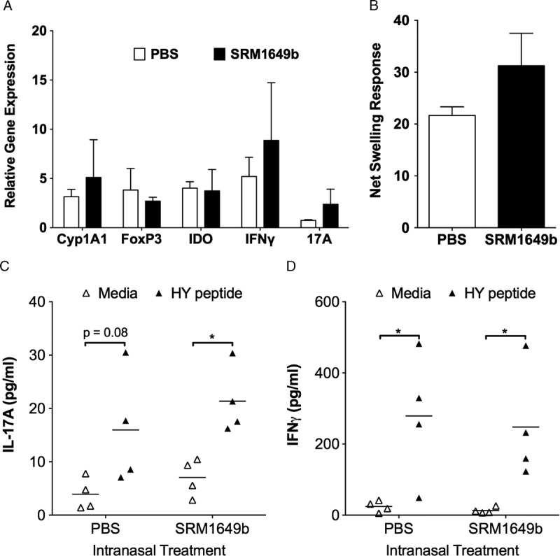
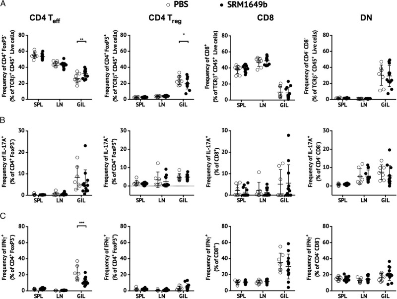
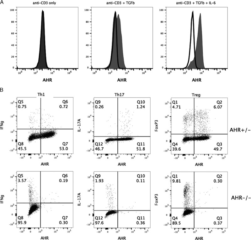
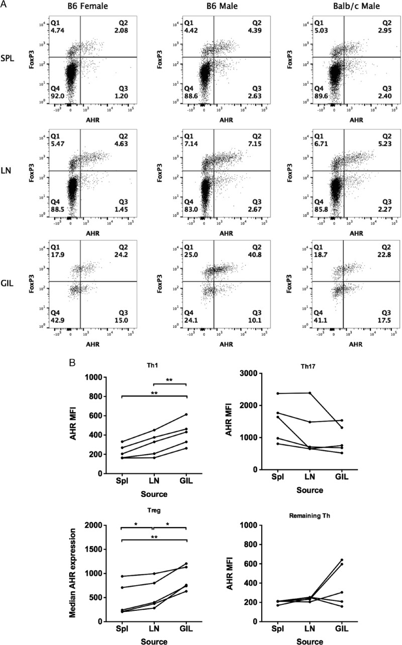
Results: Significant prolongation of male skin grafts occurred in recipient female mice treated with urban dust particles compared with controls and was found to be dependent on aryl hydrocarbon receptor (AHR) expression in the recipient mouse. T cell responses to the male histocompatibility antigen (H-Y) Dby were not altered by exposure to pollutants. A reduction in the frequency of IFNγ-producing CD4 T cells infiltrating the graft on day 7 posttransplant was observed. Flow cytometry analysis revealed that AHR expression is upregulated in IFNγ-producing CD4 T cells during immune responses in vitro and in vivo.

Conclusions: Surprisingly, inhalation of a pollutant standard was found to prolong graft survival in a minor-mismatched skin graft model in an AHR-dependent manner. One possible mechanism may be an effect on IFNγ-producing CD4 T cells responding to donor antigen. The increased expression of AHR in this CD4 T cell subset suggests that AHR ligands within the particulate matter may be directly affecting the type 1 T helper cell response in this model.


Conflict of interest statement

The authors declare no conflicts of interest.

Figures

FIGURE 1
FIGURE 1
Intranasal instillation of PM enhances IL-17A expression in lymphoid tissue. Male B6 mice were exposed to either PBS or SRM1649b, a standard PM sample of urban dust particles, by i.n. instillation every 3 days for a total of 5 doses. Twenty-four hours after the last dose, mice were killed, and SPLs and axillary LN cells were isolated. Cells were stimulated in vitro for 3 days with 1 μg/mL anti-CD3ε. Supernatants were harvested and IL-17A, and IFNγ concentration was measured by ELISA. ELISA data were analyzed by paired Student t test. **P < 0.01.
FIGURE 2
FIGURE 2
Intranasal instillation of PM prolongs allograft survival of minor but not major antigen mismatched skin grafts in an AHR-dependent manner. A, Diagram of the timing of i.n. instillation relative to skin graft. B, Male B6 mice received BALB/c skin grafts and were exposed to i.n. instillation of either PBS (n = 8; median survival time [MST] = 12.5) or SRM1649b suspended in PBS (n = 8, MST = 11), a standard PM sample of urban dust particles. Exposure of PBS or PM started 12 days before transplant and continued every 3 days for a total of 9 doses. Skin graft was performed on the day of the fifth dose. C, Female B6 mice received male B6 skin grafts and were exposed to i.n. instillation of either PBS (n = 11, MST = 25) or SRM1649b suspended in PBS (n = 16, MST = 31). D, Female AHR null mice with a B6 background mice received male B6 skin grafts and were exposed to i.n. instillation of either PBS (n = 7, MST = 21) or SRM1649b suspended in PBS (n = 10, MST = 17.5). Kaplan-Meier analysis was used to compare treatment groups.
FIGURE 3
FIGURE 3
Intranasal instillation of PM does not suppress immune responses to male antigen after engraftment of male B6 skin on female B6 recipient mice. Male B6 skin grafts were harvested from PBS- and SRM1649b-treated mice on day 18 posttransplant and processed for A, RT-PCR analysis of mRNA expression for markers of AHR activation (Cyp1A1), immune tolerance (FoxP3, indoleamine 2,3-dioxygenase), or inflammation (IFNγ, IL-17A). PCR data were normalized to actin-b expression. B, Female B6 mice received male B6 skin grafts and were exposed either PBS (n = 3) or SRM1649b suspended in PBS (n = 3) and were tested for responsiveness to the male HY antigen in a direct delayed-type hypersensitivity assay on day 18 posttransplant. Net swelling was determined by subtracting the footpad thickness at the time of injection from the footpad thickness 24 hours later. C and D, SPLs were isolated from female B6 mice treated with either PBS (n = 4) or SRM1649b (n = 4) 18 days posttransplant and stimulated in vitro for 3 days +/− Dby peptide. Culture supernatants were harvested, and IL-17A (C) and IFNγ (D) concentration were measured by ELISA. ELISA data were analyzed by paired Student t test. *P < 0.05.
FIGURE 4
FIGURE 4
Intranasal instillation of PM delays graft infiltration of IFNγ-expressing CD4 T cells after the male B6 skin to female B6 recipient skin graft model. SPLs, LNs, and GILs were harvested from PBS-treated (n = 8) and SRM1649b-treated (n = 12) female mice on day 7 posttransplant and stimulated with PMA/ionomycin in the presence of Brefeldin A for flow cytometric analysis of cytokine-expressing cells. A, percentage of TCRβ T cells that are Teff (CD4+FoxP3), Treg (CD4+FoxP3+), CD8 (CD8β+), and DN (CD4CD8β) subsets. B, percentage of IL-17A-expressing within the Teff, Treg, CD8, and DN T cell subsets. C, percentage of IFN-expressing within the Teff, Treg, CD8, and DN T cell subsets. The data were analyzed using a repeated measures 2-way ANOVA. *P < 0.05, **P < 0.01, ***P < 0.001.
FIGURE 5
FIGURE 5
Upregulation in vitro of AHR expression in CD4 T cells is not limited to the Th17 subset. A, SPLs from B6 mice (gray fill) or AHR−/− mice (black line) were stimulated with anti-CD3/CD28 +/− hTGFβ +/− mIL-6. Sixteen hours later, AHR expression within the CD4 T cell population was measured by flow cytometry. B, SPLs from AHR+/− or AHR−/− mice were stimulated with anti-CD3/CD28 +/− hTGFβ/mIL-6. Sixteen hours later, cells were stimulated for intracellular cytokine staining and, 4 hours later, stained to measure AHR expression within Th1 (IFNγ+), Th17 (IL-17A+), or Treg (FoxP3+) CD4 T cells.
FIGURE 6
FIGURE 6
AHR expression is upregulated within skin graft-infiltrating Th1 and Treg CD4 T cells. A, Female B6 mice were engrafted with female B6, male B6, or BALB/c skin without immunosuppression. Seven days later, SPLs, LN, and GIL were isolated. Cells were stained for flow cytometry to identify CD4 Teff (FoxP3) and Treg (FoxP3+) cells and to measure AHR expression. The data are representative of 2 to 5 mice per graft source. B, Female B6 mice (n = 5; pooled from 2 experiments) were engrafted with male B6 skin, and 7 days later, SPLs, LNs, and GILs were isolated. Cells were stimulated with PMA/ionomycin in the presence of Brefeldin A for flow cytometric analysis of cytokine-expressing cells. The expression of AHR was measured by median fluorescence intensity in Th1 (FoxP3, IFNγ+), Th17 (FoxP3, IL-17A+), Treg (FoxP3+), or the remaining CD4 T cells (FoxP3, IFNγ, IL-17A). To analyze the data accounting for the measurement of AHR expression in T cells from SPL, LN and GIL within the same mouse, a repeated measures 1-way ANOVA was used. *P < 0.05, **P < 0.01.

Similar articles

Cited by

References

    1. Schmidt JV, Bradfield CA. Ah receptor signaling pathways. Annu Rev Cell Dev Biol. 1996;12:55–89. - PubMed
    1. Marshall NB, Vorachek WR, Steppan LB, et al. Functional characterization and gene expression analysis of CD4+ CD25+ regulatory T cells generated in mice treated with 2,3,7,8-tetrachlorodibenzo-p-dioxin. J Immunol. 2008;181:2382–2391. - PMC - PubMed
    1. Veldhoen M, Hirota K, Westendorf AM, et al. The aryl hydrocarbon receptor links TH17-cell-mediated autoimmunity to environmental toxins. Nature. 2008;453:106–109. - PubMed
    1. Quintana FJ, Basso AS, Iglesias AH, et al. Control of T(reg) and T(H)17 cell differentiation by the aryl hydrocarbon receptor. Nature. 2008;453:65–71. - PubMed
    1. Julliard W, Fechner JH, Mezrich JD. The aryl hydrocarbon receptor meets immunology: friend or foe? A little of both. Front Immunol. 2014;5:458. - PMC - PubMed