

Kinematics in the brain: unmasking motor control strategies?
- PMID: 28573311
- PMCID: PMC5550544
- DOI: 10.1007/s00221-017-4982-8
Kinematics in the brain: unmasking motor control strategies?
Abstract
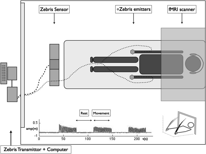
In rhythmical movement performance, our brain has to sustain movement while correcting for biological noise-induced variability. Here, we explored the functional anatomy of brain networks during voluntary rhythmical elbow flexion/extension using kinematic movement regressors in fMRI analysis to verify the interest of method to address motor control in a neurological population. We found the expected systematic activation of the primary sensorimotor network that is suggested to generate the rhythmical movement. By adding the kinematic regressors to the model, we demonstrated the potential involvement of cerebellar-frontal circuits as a function of the irregularity of the variability of the movement and the primary sensory cortex in relation to the trajectory length during task execution. We suggested that different functional brain networks were related to two different aspects of rhythmical performance: rhythmicity and error control. Concerning the latter, the partitioning between more automatic control involving cerebellar-frontal circuits versus less automatic control involving the sensory cortex seemed thereby crucial for optimal performance. Our results highlight the potential of using co-registered fine-grained kinematics and fMRI measures to interpret functional MRI activations and to potentially unmask the organisation of neural correlates during motor control.
Keywords: Error corrections; Kinematics; Motor control; Neural networks; Upper limb; fMRI.
Conflict of interest statement
Conflict of interest
The author(s) declared no potential conflict of interest with respect to the research, authorship, and/or publication of this article.
Funding
The authors declare no competing financial interests. Financial support was received from the PHRC Margaut (No. ID-RCB2010-A00596-33) and NUMEV (AN-10-LABX-20).
Ethical approval
All procedures performed in studies involving human participants were in accordance with the ethical standards of the institutional and/or national research committee and with the 1964 Helsinki Declaration and its later amendments or comparable ethical standards.
Figures


References
-
- Buma F, Kwakkel G, Ramsey N. Understanding upper limb recovery after stroke. Restor Neurol Neurosci. 2013;31:707–722. - PubMed
MeSH terms
LinkOut - more resources
Full Text Sources
Other Literature Sources
