Ridge at the medial rectus muscle insertion: A new anatomical landmark
- PMID: 28573996
- PMCID: PMC5565885
- DOI: 10.4103/ijo.IJO_214_14
Ridge at the medial rectus muscle insertion: A new anatomical landmark
Abstract
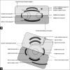
Background and aim: Rectus muscle insertions are usually linear or slight curved with the anterior convexity. While operating squint surgeries, we found a presence of ridge-like structure at the medial rectus insertion. None of the other rectus muscle insertions had such structure.
Materials and methods: Patients undergoing squint surgery were included in the study. All the patients had negative forced duction test for all the gazes and had comitant strabismus. The patients underwent surgery through the fornix route. All the squint surgeries were primary. None of the patients undergoing resurgery were included in the study. The ridge seen is actually an elevated curved structure and shows discontinuation of the actual medial rectus insertion. The measurements were taken from the superior and inferior end of the medial rectus muscle insertion.
Results: In a total of 76 medial rectus surgery (for recession or resection), we found the ridge was present in 68 (89.5%) of cases. The ridge was located at an average distance of 6.33 ± 1.5 mm inferior and 3.82 ± 0.9 mm superior to the superior and inferior point of medial rectus insertion, respectively.
Conclusion: We describe the presence, morphology, and measurements of a ridge as an anatomical landmark at medial rectus insertion.
Conflict of interest statement
There are no conflicts of interest.
Figures


References
-
- Cho HK, Shin SY. Is the insertional anatomy of rectus extraocular muscles binocularly symmetrical? Ophthalmic Res. 2010;43:179–84. - PubMed
-
- Von Noorden GK. Burian-von Noorden's binocular vision and ocular motility. 2nd ed. St. Louis: CV Mosby Co; 1980. pp. 44–452.
-
- Fink WH. Surgery of the Vertical Muscles of the Eye. 2nd ed. Springfield, Illinois: CC Thomas; 1962. pp. 90–128.
MeSH terms
LinkOut - more resources
Full Text Sources
Other Literature Sources

