Case Reports
. 2017 May;65(5):416-420.
doi: 10.4103/ijo.IJO_838_16.

Antiretinal antibody- proven autoimmune retinopathy

Affiliations
Case Reports

Antiretinal antibody- proven autoimmune retinopathy

Sharanya Abraham et al. Indian J Ophthalmol. 2017 May.

Abstract

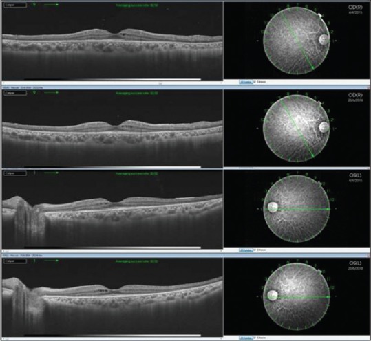
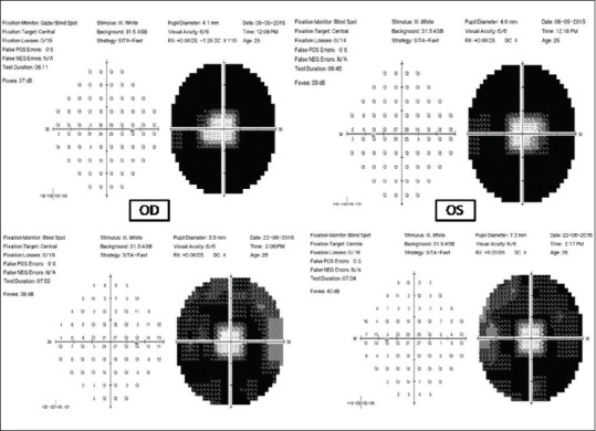
A young female presented with bilateral subacute onset of progressive decrease in night vision and reduced peripheral field of vision. The short duration and rapid progression of symptoms along with the lack of family history of night blindness prompted a diagnosis of autoimmune retinopathy (AIR). Fundus fluorescein angiography, optical coherence tomography, visual fields, and electroretinogram were suggestive of AIR. A differential diagnosis of retinitis pigmentosa (RP) was also made. Antiretinal autoantibodies were detected in the blood sample. Treatment was with oral steroids and subsequently oral immunosuppressive agents. Visual acuity was maintained, fundus examination reverted to normal, and investigations repeated at every visit were stable with improvement in visual fields. Our case suggests that AIR, if diagnosed early and treated appropriately, may have a good outcome and should be considered in patients with an atypical presentation of RP.


Conflict of interest statement

There are no conflicts of interest.

Figures

Figure 1
Figure 1
Color fundus photograph showing normal discs, attenuated vessels, retinal pigment epithelial mottling, and suspicion of cystoid macular edema
Figure 2
Figure 2
Fundus fluorescein angiogram showing perivenous patchy staining, retinal pigment epithelial defects, and disc staining
Figure 3
Figure 3
Normal fundus autofluorescence
Figure 4
Figure 4
Optical coherence tomography showing cystic spaces
Figure 5
Figure 5
Electroretinogram showing nonrecordable scotopic and photopic responses
Figure 6
Figure 6
(a) Humphrey visual fields 30-2 showing visual field constriction (b) Full-field 120 screening three-zone strategy showing visual field constriction
Figure 7
Figure 7
Serial optical coherence tomography showing reduced cystic spaces with thinning of the inner segment-outer segment junction at the macula, sparing the fovea
Figure 8
Figure 8
Humphrey visual fields 30-2 showing improved retinal sensitivity

Similar articles

Cited by

References

    1. Grange L, Dalal M, Nussenblatt RB, Sen HN. Autoimmune retinopathy. Am J Ophthalmol. 2014;157:266–72.e1. - PMC - PubMed
    1. Grewal DS, Fishman GA, Jampol LM. Autoimmune retinopathy and antiretinal antibodies: A review. Retina. 2014;34:827–45. - PubMed
    1. Ferreyra HA, Jayasundera T, Khan NW, He S, Lu Y, Heckenlively JR. Management of autoimmune retinopathies with immunosuppression. Arch Ophthalmol. 2009;127:390–7. - PubMed
    1. Choi EY, Kim M, Adamus G, Koh HJ, Lee SC. Non-paraneoplastic autoimmune retinopathy: The first case report in Korea. Yonsei Med J. 2016;57:527–31. - PMC - PubMed
    1. Heckenlively JR, Aptsiauri N, Nusinowitz S, Peng C, Hargrave PA. Investigations of antiretinal antibodies in pigmentary retinopathy and other retinal degenerations. Trans Am Ophthalmol Soc. 1996;94:179–200. - PMC - PubMed

Publication types