

A new diagnosis of Williams-Beuren syndrome in a 49-year-old man with severe bullous emphysema
- PMID: 28574231
- PMCID: PMC5509496
- DOI: 10.1002/ajmg.a.38289
A new diagnosis of Williams-Beuren syndrome in a 49-year-old man with severe bullous emphysema
Abstract
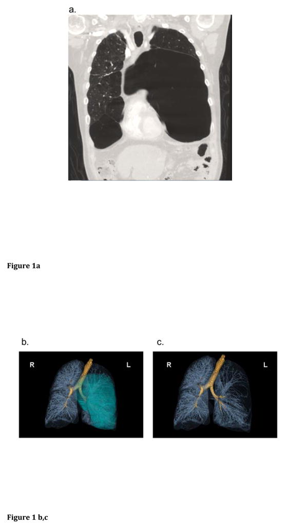
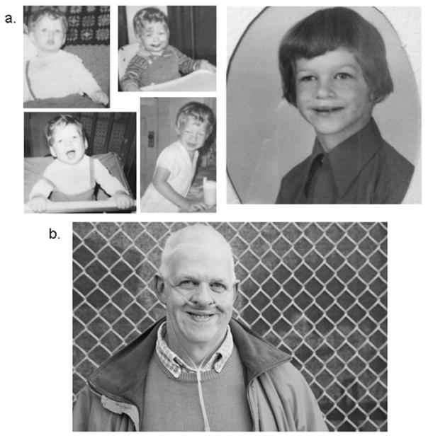
Williams-Beuren syndrome (WBS) is a chromosomal microdeletion syndrome typically presenting with intellectual disability, a unique personality, a characteristic facial appearance, and cardiovascular disease. Several clinical features of WBS are thought to be due to haploinsufficiency of elastin (ELN), as the ELN locus is included within the WBS critical region at 7q11.23. Emphysema, a disease attributed to destruction of pulmonary elastic fibers, has been reported in patients without WBS who have pathogenic variants in ELN but only once (in one patient) in WBS. Here we report a second adult WBS patient with emphysema where the diagnosis of WBS was established subsequent to the discovery of severe bullous emphysema. Haploinsufficiency of ELN likely contributed to this pulmonary manifestation of WBS. This case emphasizes the contribution of rare genetic variation in cases of severe emphysema and provides further evidence that emphysema should be considered in patients with WBS who have respiratory symptoms, as it may be under-recognized in this patient population.
Keywords: Williams-Beuren syndrome; elastin; emphysema; intellectual disability.
© 2017 Wiley Periodicals, Inc.
Figures

References
-
- Beuren AJ, Apitz J, Harmjanz D. Supravalvular Aortic Stenosis in Association with Mental Retardation and a Certain Facial Appearance. Circulation. 1962;26:1235–1240. - PubMed
-
- Boueiz A, Lutz SM, Cho MH, Hersh CP, Bowler RP, Washko GR, Halper-Stromberg E, Bakke P, Gulsvik A, Laird NM, Beaty TH, Coxson HO, Crapo JD, Silverman EK, Castaldi PJ, DeMeo DL for COPDGene and ECLIPSE investigators. Genome-Wide Association Study of the Genetic Determinants of Emphysema Distribution. Am J Respir Crit Care Med 2016 - PMC - PubMed
-
- Callewaert B, Renard M, Hucthagowder V, Albrecht B, Hausser I, Blair E, Dias C, Albino A, Wachi H, Sato F, Mecham RP, Loeys B, Coucke PJ, De Paepe A, Urban Z. New Insights into the Pathogenesis of Autosomal Dominant Cutis Laxa with Report of Five ELN Mutations. Hum Mutat. 2011;32:445. - PMC - PubMed
-
- Cherniske EM, Carpenter TO, Klaiman C, Young E, Bregman J, Insogna K, Schultz RT, Pober BR. Multisystem Study of 20 Older Adults with Williams Syndrome. Am J Med Gen Part A. 2004;131:255–264. - PubMed
Publication types
MeSH terms
Substances
Grants and funding
LinkOut - more resources
Full Text Sources
Other Literature Sources
Medical
