. 2017 Jun 2;15(6):161.
doi: 10.3390/md15060161.

Isolation, Amino Acid Sequences, and Plausible Functions of the Galacturonic Acid-Binding Egg Lectin of the Sea Hare Aplysia kurodai

Affiliations

Isolation, Amino Acid Sequences, and Plausible Functions of the Galacturonic Acid-Binding Egg Lectin of the Sea Hare Aplysia kurodai

Shoko Motohashi et al. Mar Drugs. .

Abstract

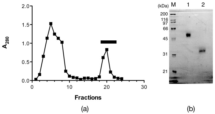
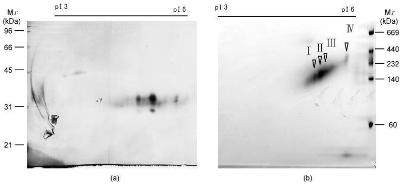
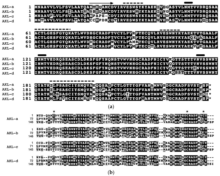
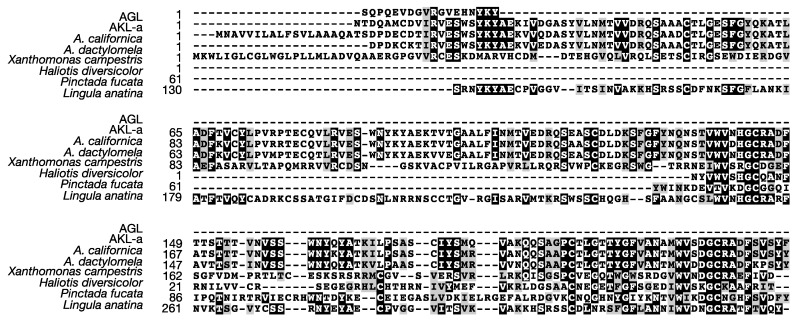
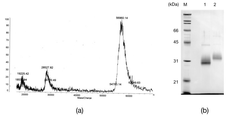
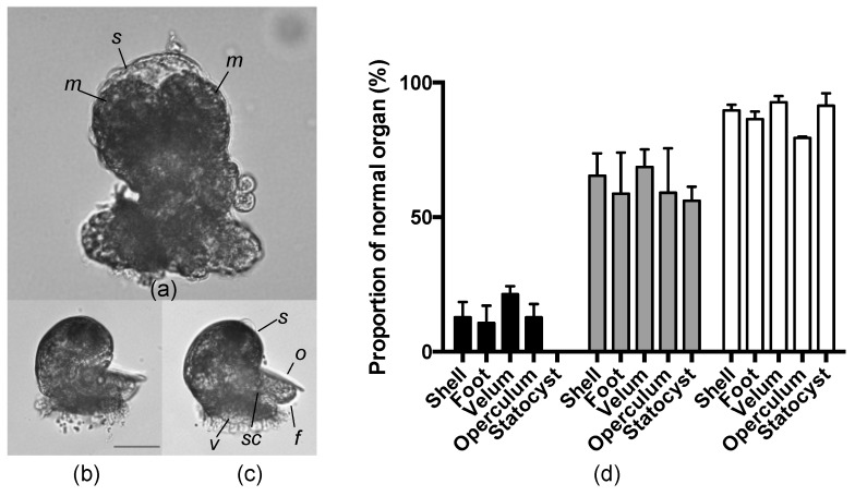
Egg lectins occur in a variety of animals ranging from mollusks to vertebrates. A few examples of molluscan egg lectins have been reported, including that of the sea hare Aplysia kurodai; however, their biological functions in the egg remain unclarified. We report the isolation, determination of primary structure, and possible functions of A.kurodai lectin (AKL) from the egg mass of A. kurodai. We obtained AKL as an inseparable mixture of isoproteins with a relative molecular mass of approximately 32 kDa by affinity purification. The hemagglutinating activity of AKL against rabbit erythrocytes was inhibited most potently by galacturonic acid and moderately by xylose. Nucleotide sequencing of corresponding cDNA obtained by rapid amplification of cDNA ends (RACE) allowed us to deduce complete amino acid sequences. The mature polypeptides consisted of 218- or 219-amino acids with three repeated domains. The amino acid sequence had similarities to hypothetical proteins of Aplysia spp., or domain DUF3011 of uncharacterized bacterial proteins. AKL is the first member of the DUF3011 family whose function, carbohydrate recognition, was revealed. Treatment of the egg with galacturonic acid, an AKL sugar inhibitor, resulted in deformation of the veliger larvae, suggesting that AKL is involved in organogenesis in the developmental stage of A. kurodai.

Keywords: Aplysia kurodai; egg lectin; embryonic development; galacturonic acid.


Conflict of interest statement

The authors declare no conflict of interest. The founding sponsors had no role in the design of the study; in the collection, analyses, or interpretation of data; in the writing of the manuscript, and in the decision to publish the results.

Figures

Figure 1
Figure 1
Affinity chromatography of Aplysia kurodai lectin (AKL). (a) affinity purification of AKL. The crude egg mass extract was applied to a d-galactose-Sepharose 6B affinity column. Unbound proteins were eluted by washing with 20 mM Tris-HCl, pH 8.0, 0.15 M NaCl (the first peak), and the bound lectin was eluted by 0.2 M d-galactose (the second peak, bar); (b) sodium dodecyl sulfate-polyacrylamide gel electrophoresis (SDS-PAGE) of purified AKL under non-reducing conditions (lane 1), reducing conditions (lane 2), and molecular standards (M).
Figure 2
Figure 2
Two-dimensional electrophoresis (2DE) of Aplysia kurodai lectin (AKL). 2DE of purified AKL under reducing conditions (a), or non-reducing conditions (b), where pI and Mr indicate isoelectric point and relative molecular mass, respectively. I, II, III and IV indicate the spots analyzed by protein sequencer.
Figure 3
Figure 3
Alignment of deduced amino acid sequences of Aplysia kurodai lectin (AKL) cDNAs. (a) the amino acid sequences deduced from the AKL cDNAs. The arrow indicates the amino terminal sequence of mature AKL. The characters with black or gray background indicate identical or similar amino acids, respectively. The thick bar indicates an N-glycosylation site. The dotted line indicates the region with a conserved sequence as determined by de novo sequencing (Table 2); (b) the tandem repeated regions of AKLs were aligned. * indicates conserved cysteine residues.
Figure 4
Figure 4
Alignment of deduced amino acid sequences of AKL-a and related proteins. The characters with black or gray background indicate identical or similar amino acids, respectively. AGL, gonad lectin of Aplysia californica (accession no. AAB24243). A hypothetical protein of the A. californica genome (accession no. XM_005101501). A galacturonic acid-binding lectin ADEL of A. dactylomela (accession no. ADP02966). A hypothetical protein of the Xanthomonas campestris genome (accession no. KIQ23628). An uncharacterized protein of Haliotis diversicolor (accession no. JU066540). Pinctada fucada, a hypothetical protein of the P. fucada gene pfu_aug2.0_1835.1_18561.t1 [25]. An uncharacterized protein of the Lingula anatina genome (accession no. XP_013393448).
Figure 5
Figure 5
Glycopeptidase F (GPF) treatment of Aplysia kurodai lectin (AKL). (a) matrix assisted laser desorption ionization-time of flight mass spectrometry (MALDI-TOF MS) for AKL; (b) Sodium dodecyl sulfate-polyacrylamide gel electrophoresis of AKL (lane 2), which digested with GPF (lane 1), and molecular standards (M).
Figure 6
Figure 6
Effect of uronic acids on the development of Aplysia kurodai larvae. Light micrographs of veliger larvae obtained from egg string treated with 50 mM GalU (a, m: macromeres, s: shell), 50 mM GlcU (b), or filtrated sea water (FSW) without sugars (c, f: foot, o: operculum, v: velum, s: shell, sc: statolith). (d) Proportion of normal organs after treatment with GalU (black bars), GlcU (gray bars), and negative control (white bars).

References

    1. Chang B.Y., Peavy T.R., Wardrip N.J., Hedrick J.L. The Xenopus laevis cortical granule lectin: cDNA cloning, developmental expression, and identification of the egg lectin family of lectins. Comp. Biochem. Physiol. Part A. 2004;137:115–129. doi: 10.1016/S1095-6433(03)00269-1. - DOI - PubMed
    1. Jimbo M., Yamashita H., Koike K., Sakai R., Kamiya H. Effects of lectin in the scleractinian coral Ctenactis echinata on symbiotic zooxanthellae. Fish. Sci. 2010;76:355–363. doi: 10.1007/s12562-009-0204-z. - DOI
    1. Koike K., Jimbo M., Sakai R., Kaeriyama M., Muramoto K., Ogata T., Maruyama T., Kamiya H. Octocoral chemical signaling selects and controls dinoflagellate symbionts. Biol. Bull. 2004;207:80–86. doi: 10.2307/1543582. - DOI - PubMed
    1. Reyes-Grajeda J.P., Moreno A., Romero A. Crystal structure of ovocleidin-17, a major protein of the calcified Gallus gallus eggshell: Implications in the calcite mineral growth pattern. J. Biol. Chem. 2004;279:40876–40881. doi: 10.1074/jbc.M406033200. - DOI - PubMed
    1. Ueda T., Nakamura Y., Smith C.M., Copits B.A., Inoue A., Ojima T., Matsunaga S., Swanson G.T., Sakai R. Isolation of novel prototype galectins from the marine ball sponge Cinachyrella sp. guided by their modulatory activity on mammalian glutamate-gated ion channels. Glycobiology. 2013;23:412–425. doi: 10.1093/glycob/cws165. - DOI - PMC - PubMed

LinkOut - more resources