. 2017 Sep;24(9):1632-1644.
doi: 10.1038/cdd.2017.91. Epub 2017 Jun 2.

NOX2-dependent ATM kinase activation dictates pro-inflammatory macrophage phenotype and improves effectiveness to radiation therapy

Affiliations

NOX2-dependent ATM kinase activation dictates pro-inflammatory macrophage phenotype and improves effectiveness to radiation therapy

Qiuji Wu et al. Cell Death Differ. 2017 Sep.

Abstract

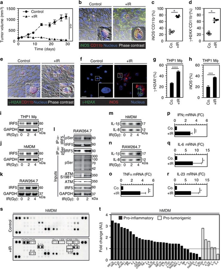
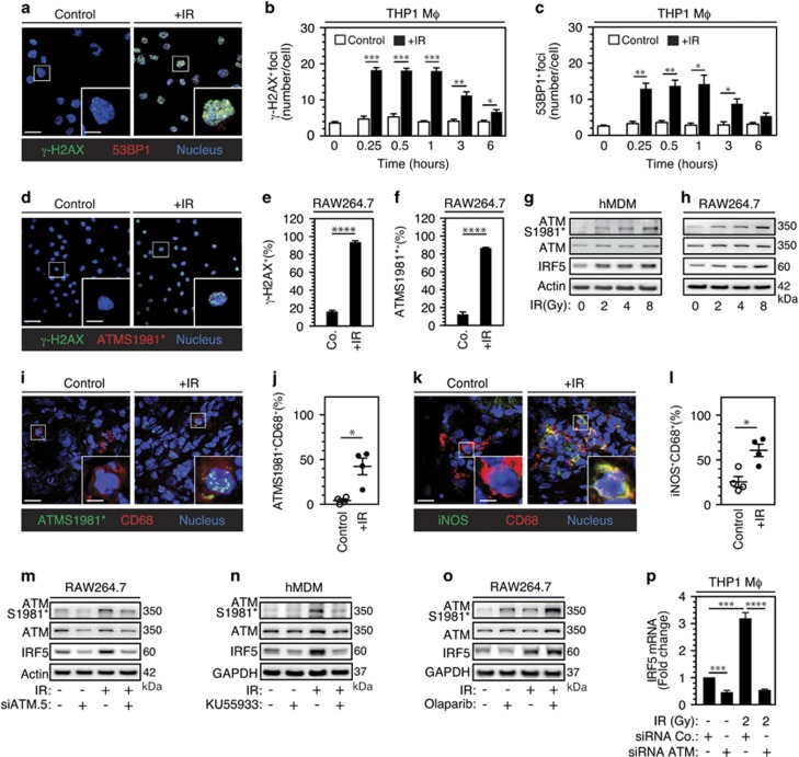
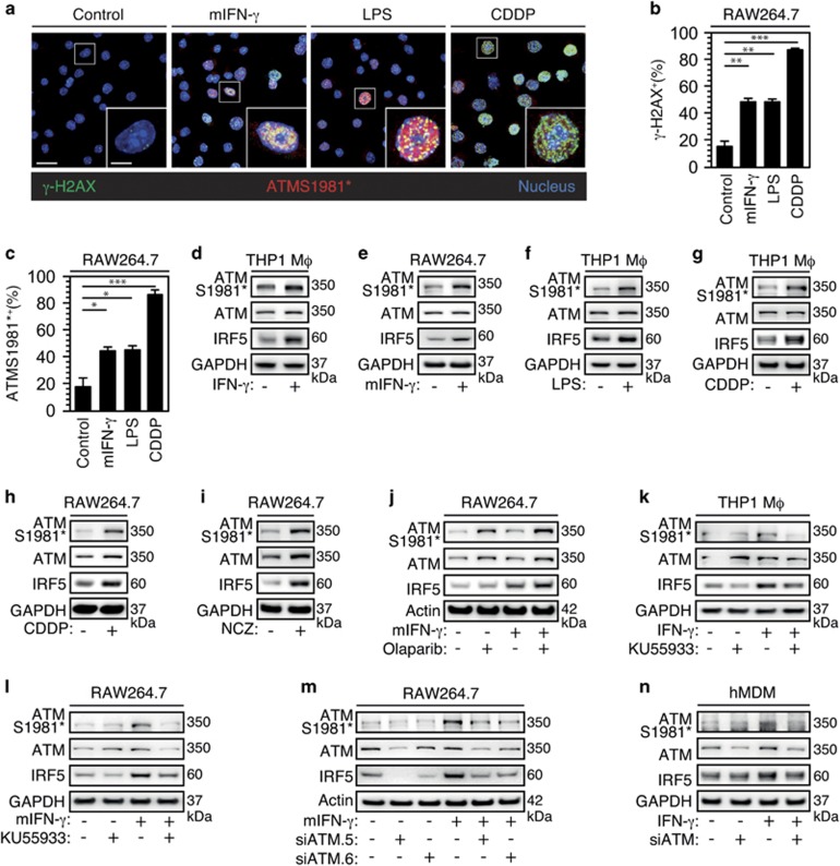
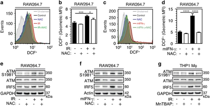
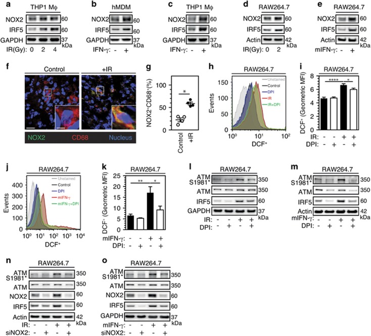
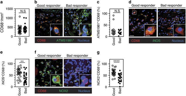
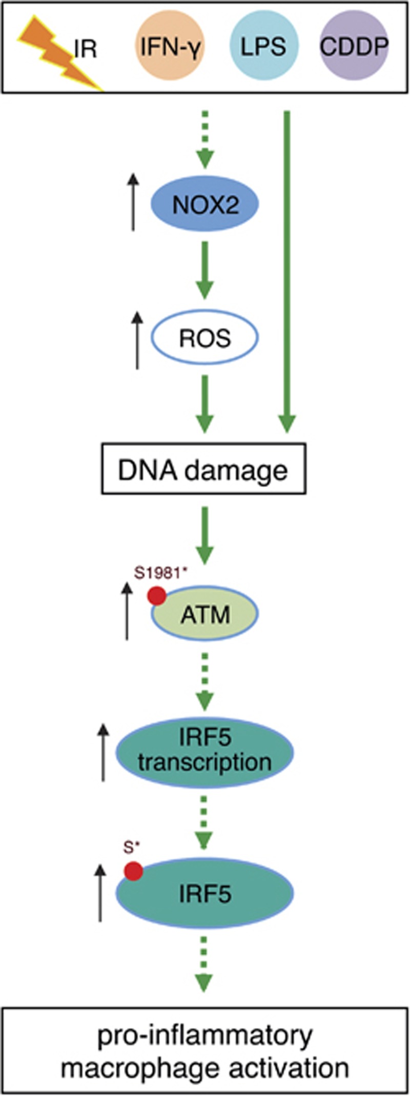
Although tumor-associated macrophages have been extensively studied in the control of response to radiotherapy, the molecular mechanisms involved in the ionizing radiation-mediated activation of macrophages remain elusive. Here we show that ionizing radiation induces the expression of interferon regulatory factor 5 (IRF5) promoting thus macrophage activation toward a pro-inflammatory phenotype. We reveal that the activation of the ataxia telangiectasia mutated (ATM) kinase is required for ionizing radiation-elicited macrophage activation, but also for macrophage reprogramming after treatments with γ-interferon, lipopolysaccharide or chemotherapeutic agent (such as cisplatin), underscoring the fact that the kinase ATM plays a central role during macrophage phenotypic switching toward a pro-inflammatory phenotype through the regulation of mRNA level and post-translational modifications of IRF5. We further demonstrate that NADPH oxidase 2 (NOX2)-dependent ROS production is upstream to ATM activation and is essential during this process. We also report that the inhibition of any component of this signaling pathway (NOX2, ROS and ATM) impairs pro-inflammatory activation of macrophages and predicts a poor tumor response to preoperative radiotherapy in locally advanced rectal cancer. Altogether, our results identify a novel signaling pathway involved in macrophage activation that may enhance the effectiveness of radiotherapy through the reprogramming of tumor-infiltrating macrophages.


Conflict of interest statement

The authors declare no conflict of interest.

Figures

Figure 1
Figure 1
Irradiation activates macrophages toward pro-inflammatory phenotype.(a) Colorectal HCT116 cells were injected subcutaneously (4 × 10⁶ cells per mouse) into immunodeficient mice and tumor growth was monitored. Results are expressed as mean value±S.E.M. P-value (δδP<0.01) was calculated by means of two-way ANOVA test. (be) Representative confocal micrographs and frequencies of iNOS+CD11b+ (b, c) or γ-H2AX+CD11b+ (d, e) tumor-associated macrophages detected in absence or after 20 Gy single-dose irradiation are shown (scale bar, 20 μm). Representative iNOS+CD11b+ or γ-H2AX+CD11b+ macrophages are shown in inserts (scale bar, 5 μm). Results are expressed as mean value±S.E.M. P-value (*P<0.05) was calculated using Mann–Whitney U-test. (fh) Representative confocal micrographs and frequencies of phorbol-12-myristate-13-acetate (PMA)-differentiated human THP1 macrophages showing γ-H2AX+ nuclear foci (f, g) or expressing iNOS (iNOS+) (f, h) in control cells or 24 h after 2 Gy irradiation are shown (scale bar, 20 μm). Representative γ-H2AX+ nuclear foci or iNOS expressing macrophages are shown in inserts (scale bar, 5 μm). Results are expressed as mean value±S.E.M. P-values (***P<0.001, ****P<0.0001) were calculated using unpaired Student’s t-test. (ik) IRF5 expression after, respectively, 96, 96 and 6 h culture of PMA-differentiated human THP1 macrophages (i), hMDM (j) or murine RAW264.7 macrophages (k) that have been irradiated (or not) with indicated doses. Representative immunoblots are shown. GAPDH is used as loading control. (l) Murine RAW264.7 macrophages that have been irradiated (or not) with 2 Gy were immunoprecipitated 6 h post irradiation for IRF5 and phopsho-serine (pSer), and analyzed for IRF5 and pSer expressions. Inputs were analyzed for IRF5, pSer, ATMS1981*, ATM and GAPDH. (m, n) Detection of IL-1β and IL-8 release in the supernatants of hMDM (m) or murine RAW264.7 macrophages (n) that have been irradiated (or not) with indicated doses. Representative immunoblots are shown. (or) TNFα, IFNγ, IL-6 and IL-23 mRNA expressions on PMA-differentiated THP1 macrophages that have been irradiated (or not) with 2 Gy were determined by quantitative real-time PCR. Results are expressed as mean value±S.E.M. and represented as fold change as compared to controls. P-values (*P<0.05, **P<0.01) were calculated using unpaired Student’s t-test. (s, t) Detection of cytokine secretion in the supernatants of hMDMs that have been treated (or not) with 4 Gy irradiation. Array images were captured following 1–10 min exposures to peroxidase substrate (s). Relative levels of cytokines detected in the supernatants of irradiated macrophages as compared to those detected in non-irradiated macrophages are revealed as fold change of arbitrary units. Pro-inflammatory and pro-tumorigenic cytokines and chemokines are indicated (t). Data are representative of three independent experiments performed with primary human macrophages obtained from three healthy representative donors
Figure 2
Figure 2
ATM activation controls IRF5 transcriptional expression and IR-induced pro-inflammatory macrophage phenotype.(a) Representative confocal micrographs of phorbol-12-myristate-13-acetate (PMA)- differentiated human THP1 macrophages showing γ-H2AX+ or 53BP+ foci following 2 Gy single-dose irradiation are shown (scale bar, 20 μm). Scale bar of inserts is 5 μm. (b, c) Frequencies of γ-H2AX+ (b) or 53BP+ (c) nuclear foci in PMA-differentiated human THP1 macrophages after 2 Gy single-dose irradiation are shown at indicated times. (df) Representative confocal micrographs and frequencies of murine RAW264.7 macrophages showing γ-H2AX+ nuclear foci (d, e) or AMTS1981* phosphorylation (ATMS1981*+) (d, f), in control cells or 1 h after 2 Gy single-dose irradiation are shown (scale bar, 20 μm). Representative γ-H2AX+ nuclear foci and ATMS1981*+ macrophages are shown in inserts (scale bar, 5 μm). Results are expressed as mean value±S.E.M. P-values (*P<0.05, **P<0.01, ***P<0.001, ****P<0.0001) were calculated using unpaired Student’s t-test. (g, h) ATMS1981*, ATM and IRF5 expression after, respectively, 96 and 6 h culture of hMDM (g) or murine RAW264.7 macrophages (h) that have been irradiated (or not) with indicated doses are determined. Representative immunoblots are shown. Actin is used as loading control. (il) Representative confocal micrographs and frequencies of ATMS1981*+CD68+ (i, j) or iNOS+CD68+ (k, l) macrophages that have been detected in absence or after 45 Gy total dose of fractionated irradiation on tumor samples obtained from locally advanced rectal cancer patients are shown (scale bar, 20 μm; scale bar of insert, 5 μm). Results are expressed as mean value±S.E.M. P-value (*P<0.05) was calculated using Mann–Whitney U-test. (m, n) ATMS1981*, ATM and IRF5 expression after, respectively, 6 and 96 h culture of murine RAW264.7 macrophages that have been depleted for ATM (m) or hMDM that have been treated with 20 μM of KU55933 (n) and irradiated (or not) with 2 Gy (m) or 4 Gy (n) are shown. Representative immunoblots are shown. GAPDH (or actin) is used as loading control. (o) ATMS1981*, ATM and IRF5 expression after 6 h culture of murine RAW264.7 macrophages that have been treated with 10 μM of Olaparib and irradiated (or not) with 2 Gy are shown. Representative immunoblots are shown. GAPDH is used as loading control. (p) IRF5 mRNA expression on PMA-differentiated human THP1 macrophages that have been depleted for ATM and irradiated (or not) with 2 Gy was determined by quantitative real-time PCR. Results are expressed as mean value±S.E.M. and represented as fold change as compared to controls. P-values (***P<0.001 and ****P<0.0001) were calculated using one-way ANOVA test. Quantification of western blot bands are shown in Supplementary Figure 4
Figure 3
Figure 3
Classical macrophage activation is dependent on ATM. (ac) Representative confocal micrographs and frequencies of murine RAW264.7 macrophages showing γ-H2AX+ nuclear foci (a, b) or ATMS1981* phosphorylation (ATMS1981*+) (a, c) in control cells or after 24 h treatments with 20 ng/ml of recombinant murine IFN-γ (mIFN-γ), 100 ng/ml of lipopolysaccharide (LPS) or 10 μM of cisplatinium (CDDP) are shown (scale bar, 20 μm). Representative macrophages with ATMS1981*+ and γ-H2AX+ nuclear foci are shown in inserts (scale bar, 5 μm). Results are expressed as mean value±S.E.M. P-values (*P<0.05, **P<0.01, ***P<0.001) were calculated using unpaired Student’s t-test. (di) ATMS1981*, ATM and IRF5 expressions after 24 h culture of PMA-differentiated human THP1 macrophages (d, f, g) or murine RAW264.7 macrophages (e, h, i) that have been treated (or not) with 20 ng/ml of recombinant human IFN-γ (IFN-γ) (d), 20 ng/ml of recombinant murine IFN-γ (mIFN-γ) (e), 100 ng/ml of LPS (f), 10 μM of CDDP (g, h) or 200 ng/ml of neocarzinostatin (NCZ) (i) are determined. Representative immunoblots are shown. GAPDH is used as loading control. (jn) ATMS1981*, ATM and IRF5 expressions after, respectively, 24 h culture of murine RAW264.7 macrophages (j, l, m), PMA-differentiated human THP1 macrophages (κ) or hMDMs (n) that have been incubated with 10 μM of Olaparib (j), with 20 μM of KU55933 (k, l) or depleted for ATM (m, n) and treated (or not) with 20 ng/ml mIFN-γ (for RAW264.7 macrophages) (j, l, m), 20 ng/ml human IFN-γ (for PMA-differentiated human THP1 macrophages) (κ) or 4 μg/ml of human IFN-γ (for hMDM) (n) are evaluated. Representative immunoblots are shown. Actin (or GAPDH) is used as loading control. Quantification of western blot bands is shown in Supplementary Figure 4
Figure 4
Figure 4
Reactive oxygen species are involved in IR-induced pro-inflammatory macrophage activation.(ad) Murine RAW264.7 macrophages treated with 1 μg/ml of NAC were stimulated with 2 Gy single-dose irradiation (a, b) or 20 ng/ml mIFN-γ (c, d), stained with H2DCFDA and analyzed by flow cytometry. Representative flow cytometry analysis and quantifications of geometric mean fluorescence intensity (MFI) are shown. Data are presented as means±S.E.M. in b and d panels. Significances are *P<0.05, ***P<0.001 and ****P<0.0001, and were obtained using one-way ANOVA test. (e, f) ATMS1981*, ATM and IRF5 expressions after, respectively, 6 and 24 h culture of murine RAW264.7 macrophages that have been incubated with 1 μg/ml of NAC and irradiated with 2 Gy single dose (e) or treated with 20 ng/ml mIFN-γ (f) were determined. Representative immunoblots are shown. GAPDH (or actin) is used as loading control. (g) ATMS1981*, ATM and IRF5 expressions after 48 h culture of PMA-differentiated human THP1 macrophages that have been incubated with 10 μM of MnTBAP and irradiated with 8 Gy single dose were determined. Representative immunoblots are shown. GAPDH is used as loading control. Quantification of western blot bands are shown in Supplementary Figure 5
Figure 5
Figure 5
NOX2-dependent ROS production is involved in the pro-inflammatory macrophage activation. (ae) NOX2 and IRF5 expressions after, respectively, 96 and 6 h culture of PMA-differentiated human THP1 macrophages (a) or murine RAW264.7 macrophages (d) that have been irradiated (or not) with indicated doses (a and d); or 24 h culture of hMDM with 4 μg/ml of human IFN-γ (b), PMA-differentiated human THP1 macrophages with 20 ng/ml of human IFN-γ (c) or murine RAW264.7 macrophages with 20 ng/ml of mIFN-γ (e) were determined. Representative immunoblots are shown. GAPDH and actin were used as loading control. (f, g) Representative confocal micrographs and frequencies of NOX2+CD68+ tumor-associated macrophages detected in absence or after 45 Gy total dose of fractionated irradiation on tumor samples obtained from locally advanced rectal cancer patients are shown (scale bar, 20 μm; scale bar of insert, 5 μm). Results are expressed as mean value±S.E.M. P-value (*P<0.05) was calculated using Mann–Whitney U-test. (hk) Murine RAW264.7 macrophages treated with 200 nM of DPI and irradiated with 2 Gy single dose (h, i) or stimulated with 20 ng/ml mIFN-γ (j, k) stained with H2DCFDA and analyzed by flow cytometry. Representative flow cytometry analysis and quantifications of geometric MFI are shown. Data are presented as means±S.E.M. in i and k panels. Significances are *P<0.05, **P<0.01 and ****P<0.0001, and were obtained using one-way ANOVA test. (lo) ATMS1981*, ATM and IRF5 expressions after, respectively, 6 and 24 h culture of murine RAW264.7 macrophages that have been incubated with 200 nM of DPI (l, m) or depleted for NOX2 (n, o) and irradiated with 2 Gy single dose (l, n) or treated with 20 ng/ml mIFN-γ (m, o) were determined. Representative immunoblots are shown. GAPDH and actin were used as loading control. Quantification of western blot bands are shown in Supplementary Figure 6
Figure 6
Figure 6
The perturbation of NOX2/ATM-dependent signaling pathway is associated with poor tumor response to radiation therapy.(a) Densities of CD68+ tumor-infiltrating macrophages detected on biopsies of human rectal tumor samples from good responders (n=29) and bad responders (n=27) to neoadjuvant radiation therapy were analyzed. Data are presented as means±S.E.M. (b) Representative confocal micrographs and frequencies of ATMS1981*+CD68+ (b, c), iNOS+CD68+ (d, e) or NOX2+CD68+ (f, g) tumor-associated macrophages detected in good responders (n=29) and bad responders (n=27) to neoadjuvant radiation therapy are shown (scale bar, 20 μm). Representative ATMS1981*+CD68+, iNOS+CD68+ or NOX2+CD68+ macrophages are shown in inserts (scale bar, 5 μm). Results are expressed as mean value±S.E.M. P-values (**P<0.01 and ***P<0.001) were calculated using Mann–Whitney U-test
Figure 7
Figure 7
Proposed model for the roles of NOX2 and ATM activations in pro-inflammatory macrophage activation

Similar articles

Cited by

References

    1. Azzam EI, Jay-Gerin JP, Pain D. Ionizing radiation-induced metabolic oxidative stress and prolonged cell injury. Cancer Lett 2012; 327: 48–60. - PMC - PubMed
    1. Hekim N, Cetin Z, Nikitaki Z, Cort A, Saygili EI. Radiation triggering immune response and inflammation. Cancer Lett 2015; 368: 156–163. - PubMed
    1. Park B, Yee C, Lee KM. The effect of radiation on the immune response to cancers. Int J Mol Sci 2014; 15: 927–943. - PMC - PubMed
    1. Kroemer G, Galluzzi L, Kepp O, Zitvogel L. Immunogenic cell death in cancer therapy. Annu Rev Immunol 2013; 31: 51–72. - PubMed
    1. Ghiringhelli F, Apetoh L, Tesniere A, Aymeric L, Ma Y, Ortiz C et al. Activation of the NLRP3 inflammasome in dendritic cells induces IL-1beta-dependent adaptive immunity against tumors. Nat Med 2009; 15: 1170–1178. - PubMed

Substances