

Characterization of the release and biological significance of cell-free DNA from breast cancer cell lines
- PMID: 28574818
- PMCID: PMC5522137
- DOI: 10.18632/oncotarget.17858
Characterization of the release and biological significance of cell-free DNA from breast cancer cell lines
Abstract
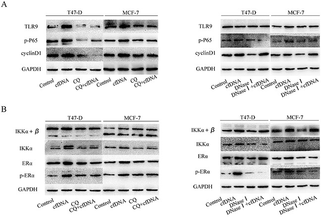
In breast cancer, cell-free DNA (cfDNA) has been proven to be a diagnostic and prognostic biomarker. However, there have been few studies on the origin and biological significance of cfDNA. In this study, we assessed the release pattern of cfDNA from breast cancer cell lines under different culture conditions and investigated the biological significance of cfDNA. The cfDNA concentration increased rapidly (6 h) after passage, decreased gradually, and was then maintained at a relatively stable level after 24 h. In addition, the cfDNA concentration did not correlate with the amount of apoptotic and necrotic cells. Interestingly, if more cells were in the G1 phase, more cfDNA was detected (p < 0.01) and the cfDNA concentration correlated positively with the percent of cells in the G1 phase (p < 0.05). We observed that cells could release cfDNA actively, but not exclusively, via exosomes. Furthermore, we showed that cfDNA could stimulate hormone receptor-positive breast cancer cell proliferation by activating the TLR9-NF-κB-cyclin D1 pathway. In conclusion, cfDNA is released from breast cancer mainly by active secretion, and cfDNA could stimulate proliferation of breast cancer cells.
Keywords: biological significance; breast cancer; cell-free DNA; circulating tumor DNA.
Conflict of interest statement
The authors declare that they have no conflict of interest.
Figures


Similar articles
-
Circulating cell-free DNA as a prognostic and predictive biomarker in non-small cell lung cancer.Oncotarget. 2016 Jul 12;7(28):44583-44595. doi: 10.18632/oncotarget.10069. Oncotarget. 2016. PMID: 27323821 Free PMC article.
-
Cell-free DNA derived from cancer cells facilitates tumor malignancy through Toll-like receptor 9 signaling-triggered interleukin-8 secretion in colorectal cancer.Acta Biochim Biophys Sin (Shanghai). 2018 Oct 1;50(10):1007-1017. doi: 10.1093/abbs/gmy104. Acta Biochim Biophys Sin (Shanghai). 2018. PMID: 30239551
-
[Characteristics and diagnostic applications of circulating cell-free DNA in colorectal cancer].Orv Hetil. 2019 Jul;160(30):1167-1177. doi: 10.1556/650.2019.31486. Orv Hetil. 2019. PMID: 31327245 Review. Hungarian.
-
Circulating free DNA integrity and concentration as independent prognostic markers in metastatic breast cancer.Breast Cancer Res Treat. 2018 May;169(1):69-82. doi: 10.1007/s10549-018-4666-5. Epub 2018 Jan 16. Breast Cancer Res Treat. 2018. PMID: 29340881
-
Putative Origins of Cell-Free DNA in Humans: A Review of Active and Passive Nucleic Acid Release Mechanisms.Int J Mol Sci. 2020 Oct 29;21(21):8062. doi: 10.3390/ijms21218062. Int J Mol Sci. 2020. PMID: 33137955 Free PMC article. Review.
Cited by
-
Serial profiling of cell-free DNA and nucleosome histone modifications in cell cultures.Sci Rep. 2021 May 4;11(1):9460. doi: 10.1038/s41598-021-88866-5. Sci Rep. 2021. PMID: 33947882 Free PMC article.
-
Tumor-derived exosomal HMGB1 promotes esophageal squamous cell carcinoma progression through inducing PD1+ TAM expansion.Oncogenesis. 2019 Feb 22;8(3):17. doi: 10.1038/s41389-019-0126-2. Oncogenesis. 2019. PMID: 30796203 Free PMC article.
-
Multidimensional fragmentomic profiling of cell-free DNA released from patient-derived organoids.Hum Genomics. 2023 Oct 28;17(1):96. doi: 10.1186/s40246-023-00533-0. Hum Genomics. 2023. PMID: 37898819 Free PMC article.
-
Non-small cell lung carcinoma (NSCLC): Implications on molecular pathology and advances in early diagnostics and therapeutics.Genes Dis. 2022 Aug 23;10(3):960-989. doi: 10.1016/j.gendis.2022.07.023. eCollection 2023 May. Genes Dis. 2022. PMID: 37396553 Free PMC article. Review.
-
DNA in extracellular vesicles: biological and clinical aspects.Mol Oncol. 2021 Jun;15(6):1701-1714. doi: 10.1002/1878-0261.12777. Epub 2020 Aug 19. Mol Oncol. 2021. PMID: 32767659 Free PMC article. Review.
References
-
- Spellman PT, Gray JW. Detecting cancer by monitoring circulating tumor DNA. Nature Med. 2014;20:474–475. - PubMed
-
- Dawson SJ, Tsui DW, Murtaza M, Biggs H, Rueda OM, Chin SF, Dunning MJ, Gale D, Forshew T, Mahler-Araujo B, Rajan S, Humphray S, Becq J, et al. Analysis of circulating tumor DNA to monitor metastatic breast cancer. N Engl J Med. 2013;368:1199–1209. - PubMed
-
- Schwarzenbach H, Hoon DS, Pantel K. Cell-free nucleic acids as biomarkers in cancer patients. Nat Rev Cancer. 2011;11:426–437. - PubMed
MeSH terms
Substances
LinkOut - more resources
Full Text Sources
Other Literature Sources
Medical
Research Materials
