. 2017 Jun 1;96(6):1197-1209.
doi: 10.1093/biolre/iox047.

Maternal RNA regulates Aurora C kinase during mouse oocyte maturation in a translation-independent fashion

Affiliations

Maternal RNA regulates Aurora C kinase during mouse oocyte maturation in a translation-independent fashion

Ahmed Z Balboula et al. Biol Reprod. .

Abstract

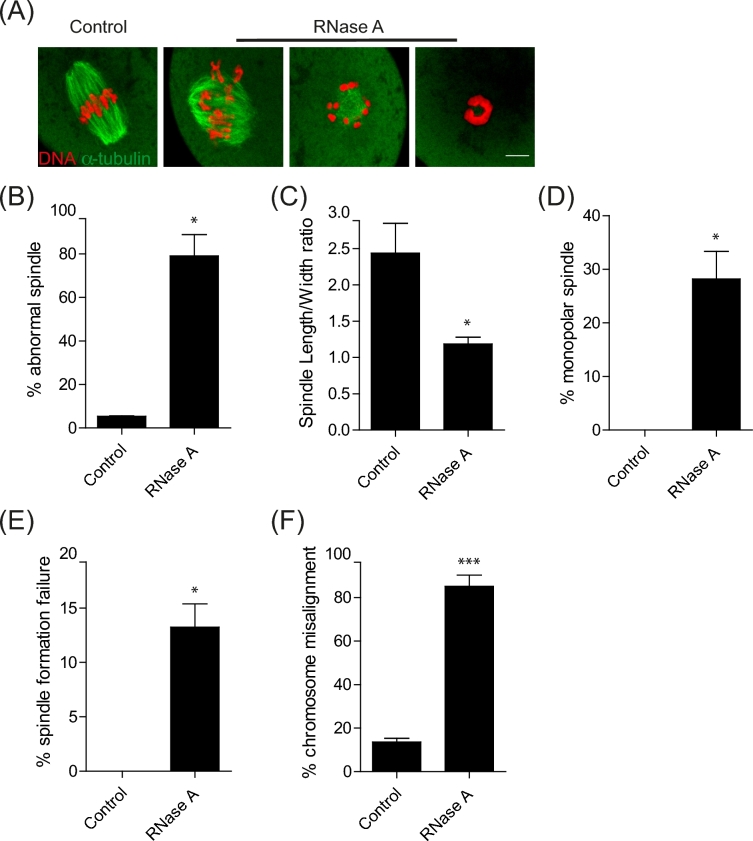
During oocyte meiotic maturation, Aurora kinase C (AURKC) is required to accomplish many critical functions including destabilizing erroneous kinetochore-microtubule (K-MT)attachments and regulating bipolar spindle assembly. How localized activity of AURKC is regulated in mammalian oocytes, however, is not fully understood. Female gametes from many species, including mouse, contain stores of maternal transcripts that are required for downstream developmental events. We show here that depletion of maternal RNA in mouse oocytes resulted in impaired meiotic progression, increased incidence of chromosome misalignment and abnormal spindle formation at metaphase I (Met I), and cytokinesis defects. Importantly, depletion of maternal RNA perturbed the localization and activity of AURKC within the chromosomal passenger complex (CPC). These perturbations were not observed when translation was inhibited by cycloheximide (CHX) treatment. These results demonstrate a translation-independent function of maternal RNA to regulate AURKC-CPC function in mouse oocytes.

Keywords: Aurora kinase C; chromosomal passenger complex; maternal RNA; meiosis; mouse oocyte.


Figures

Figure 1.
Figure 1.
Depletion of maternal RNA impairs oocyte maturation. Full-grown prophase I-arrested oocytes were injected with PBS or RNase A followed by maturation in vitro for 16 h. (A) First polar body extrusion (PBE) was scored to assess meiotic progression. (B) Quantification of cytokinesis defects based on live cell imaging (n = 21, 26). (C) Representative live cell images showing retraction of the extruded polar body in RNA-depleted oocytes; scale bar represents 100 μm. White arrowheads in the lower panel indicates chromatin aggregation; scale bar, 10 μm. (D) Confocal microscopy images of Met II eggs stained with an anti-α-tubulin antibody to label spindle (green) and DAPI to label DNA (red). Representative images are shown. (E) Quantification of chromosomal collapse phenotype. The experiments were carried out at least three times and the total numbers of oocytes examined are indicated above the graph bars. The data are expressed as mean ± SEM, and Student t-test was used to analyze the data except (A) where one-way ANOVA was used to analyze the data. Values with asterisks vary significantly, *P < 0.05, **P < 0.01.
Figure 2.
Figure 2.
Maternal RNA perturbs bipolar spindle assembly. Full-grown, prophase I-arrested oocytes were injected with PBS or RNase A followed by in vitro maturation to Met I. (A) Met I oocytes were fixed and stained with an anti-α-tubulin antibody to label spindle microtubules (green) and DAPI to label DNA (red) followed by confocal imaging. Representative images are shown. Scale bar represents 10 μm. (B) Quantification of abnormal spindle morphology (C) Quantification of spindle length/width ratio. (D) Quantification of the number of oocytes with a monopolar spindle. (E) Quantifications of oocytes that failed to form a spindle. (F) Quantification of the number of oocytes with misaligned chromosomes. The experiment was carried out three times, and the total number of oocytes examined were 30 and 34 oocytes in the control and RNase A groups, respectively. The data are expressed as mean ± SEM and Student t-test was used to analyze the data. Values with asterisks vary significantly, *P < 0.05, ***P < 0.001.
Figure 3.
Figure 3.
Maternal RNA perturbs AURKC localization during oocyte maturation. Full-grown prophase I-arrested oocytes were injected with PBS or RNase A followed by in vitro maturation. (A) Met I oocytes (6 h) were fixed and immunostained with an anti-AURKC antibody (green in merge). DNA was detected with DAPI (blue in merge). (B) Corresponding quantification of oocytes with localized AURKC in (A). (C) Telophase I oocytes were fixed and immunostained with an anti-AURKC antibody (red in merge) and an anti-α-tubulin antibody (green in merge). DNA was labeled with DAPI (blue in merge). The scale bar represents 10 μm, and representative images are shown. The experiments were carried out two times and the total number of oocytes examined were 20 and 24 oocytes in the control and RNase A groups, respectively. The data are expressed as mean ± SEM, and Student t-test was used to analyze the data. Values with asterisks vary significantly, ***P < 0.001.
Figure 4.
Figure 4.
Maternal RNA perturbs CPC localization and activity during oocyte maturation. Full-grown prophase I-arrested oocytes were injected with PBS or RNase A followed by in vitro maturation to Met I. Met I oocytes (6 h) were fixed and immunostained (green in merge) with an anti-survivin antibody (A), anti-pINCENP antibody (C), and anti-H3pS10 antibody (E). DNA was detected with DAPI (blue in merge). Representative images are shown, scale bar represents 10 μm. (B), (D) and (F) Corresponding quantifications of fluorescence intensities of (A), (C) and (E), respectively. The experiments were carried out two times and the total numbers of examined oocytes are indicated above the graph bars. The data are expressed as mean ± SEM, and Student t-test was used to analyze the data. Values with asterisks vary significantly, *P < 0.05, **P < 0.01, ***P < 0.001.
Figure 5.
Figure 5.
Translation of maternal RNA is not required for AURKC localization and activity. Full-grown prophase I-arrested oocytes either noninjected or injected with Gfp cRNA were in vitro matured in the presence of PBS or 10 μg/ml cycloheximide for 6 h. Met I oocytes were fixed and examined for GFP expression (A; green in merge) or immunostained (red in merge) with an anti-AURKC antibody (C), or anti-H3pS10 antibody (E). DNA was detected with DAPI (blue in merge). The scale bar represents 50 μm for (A) and 10 μm for (C and E). Representative images are shown. The experiments were carried out two times and the total numbers of examined oocytes are indicated above the graph bars. (B), (D), and (F) Corresponding fluorescence intensity quantifications for (A), (C), and (E), respectively. The data are expressed as mean ± SEM, and Student t-test was used to analyze the data.
Figure 6.
Figure 6.
Regulation of AURKC and meiotic spindle by maternal RNA during oocyte maturation is independent of translation. Full-grown prophase I-arrested oocytes were matured in vitro for 6 h (Met I) or 14 h (Met II). Met I and Met II oocytes were injected with PBS or RNase A followed by incubation in the same maturation condition for an additional 2 h. Met I (A-C) and Met II (D-F) injected oocytes were fixed and immunostained with an anti-AURKC antibody (red in merge) and an anti-α-tubulin antibody (green in merge). DNA was labeled with DAPI (blue in merge). The scale bar represents 10 μm, and representative images are shown. (B, C) Corresponding quantifications of oocytes in A. (E, F) Corresponding quantifications of eggs from D. The experiments were carried out two times and the total numbers of examined oocytes are indicated above the graph bars. The data are expressed as mean ± SEM, and Student t-test was used to analyze the data. Values with asterisks vary significantly, *P < 0.05, ***P < 0.001.

References

    1. Ditchfield C, Johnson VL, Tighe A, Ellston R, Haworth C, Johnson T, Mortlock A, Keen N, Taylor SS. Aurora B couples chromosome alignment with anaphase by targeting BubR1, Mad2, and Cenp-E to kinetochores. J Cell Biol 2003; 161:267–280. - PMC - PubMed
    1. Birkenfeld J, Nalbant P, Bohl BP, Pertz O, Hahn KM, Bokoch GM. GEF-H1 modulates localized RhoA activation during cytokinesis under the control of mitotic kinases. Dev Cell 2007; 12:699–712. - PMC - PubMed
    1. Jambhekar A, Emerman AB, Schweidenback CT, Blower MD. RNA stimulates Aurora B kinase activity during mitosis. PLoS One 2014; 9:e100748. - PMC - PubMed
    1. Jelluma N, Brenkman AB, van den Broek NJ, Cruijsen CW, van Osch MH, Lens SM, Medema RH, Kops GJ. Mps1 phosphorylates Borealin to control Aurora B activity and chromosome alignment. Cell 2008; 132:233–246. - PubMed
    1. Shuda K, Schindler K, Ma J, Schultz RM, Donovan PJ. Aurora kinase B modulates chromosome alignment in mouse oocytes. Mol Reprod Dev 2009; 76:1094–1105. - PMC - PubMed