. 2017 Jul 27;45(13):8026-8045.
doi: 10.1093/nar/gkx477.

Major hnRNP proteins act as general TDP-43 functional modifiers both in Drosophila and human neuronal cells

Affiliations

Major hnRNP proteins act as general TDP-43 functional modifiers both in Drosophila and human neuronal cells

Chiara Appocher et al. Nucleic Acids Res. .

Abstract

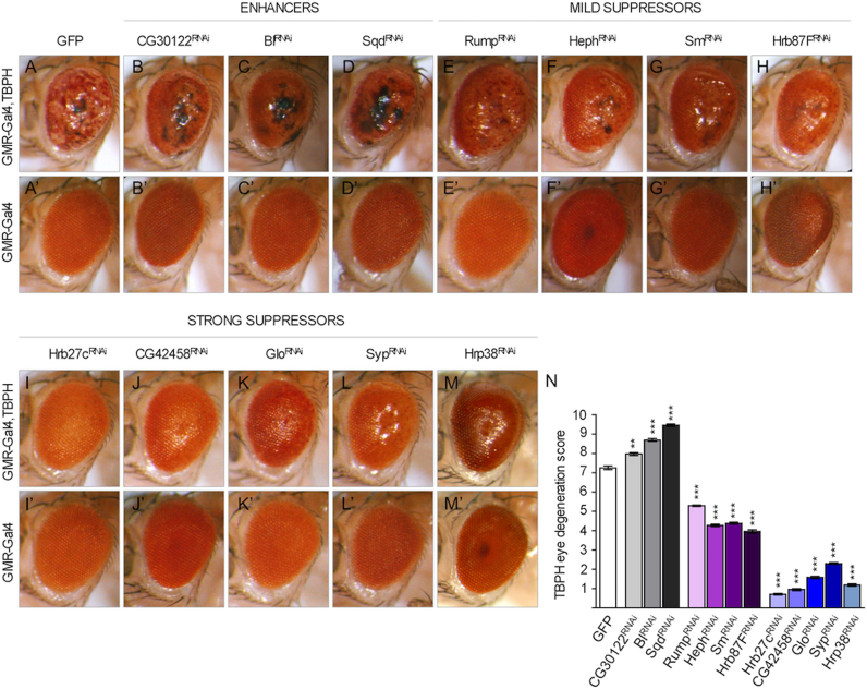
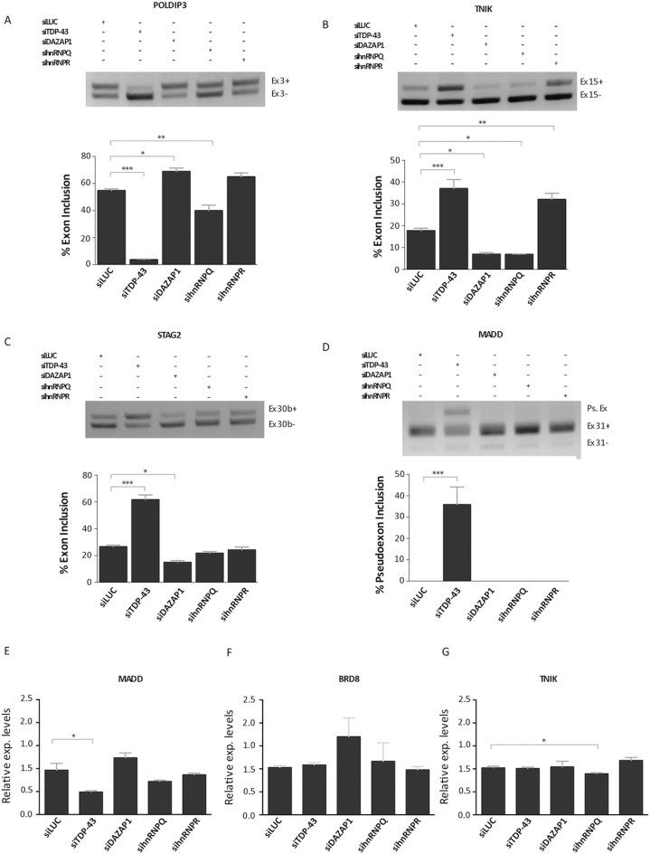
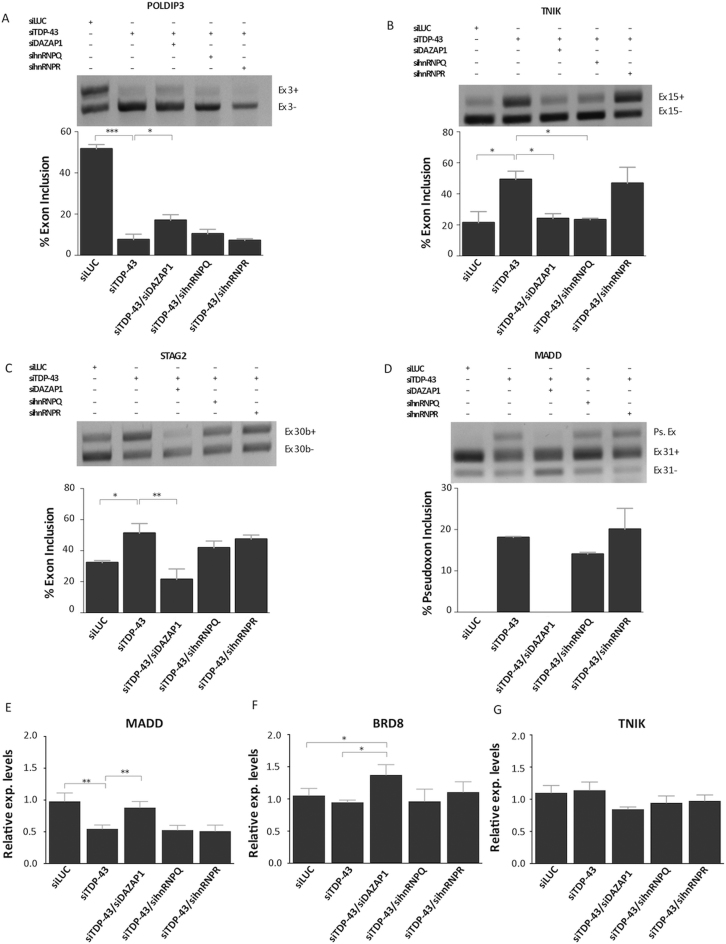
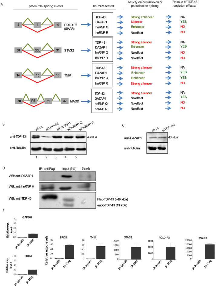
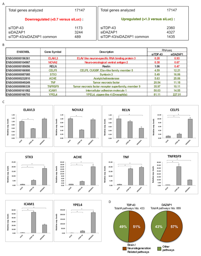
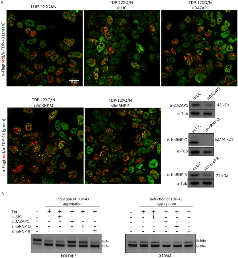
Nuclear factor TDP-43 is known to play an important role in several neurodegenerative pathologies. In general, TDP-43 is an abundant protein within the eukaryotic nucleus that binds to many coding and non-coding RNAs and influence their processing. Using Drosophila, we have performed a functional screening to establish the ability of major hnRNP proteins to affect TDP-43 overexpression/depletion phenotypes. Interestingly, we observed that lowering hnRNP and TDP-43 expression has a generally harmful effect on flies locomotor abilities. In parallel, our study has also identified a distinct set of hnRNPs that is capable of powerfully rescuing TDP-43 toxicity in the fly eye (Hrb27c, CG42458, Glo and Syp). Most importantly, removing the human orthologs of Hrb27c (DAZAP1) in human neuronal cell lines can correct several pre-mRNA splicing events altered by TDP-43 depletion. Moreover, using RNA sequencing analysis we show that DAZAP1 and TDP-43 can co-regulate an extensive number of biological processes and molecular functions potentially important for the neuron/motor neuron pathophysiology. Our results suggest that changes in hnRNP expression levels can significantly modulate TDP-43 functions and affect pathological outcomes.


Figures

Figure 1.
Figure 1.
RNAi-mediated disruption of hnRNP candidate genes in Drosophila eyes. (A–M) RNAi-mediated knockdown of hnRNPs alters phenotype of TBPH gain-of-function eyes. Compared the control fly expressing TBPH (A) GMR-Gal4, TBPH/GFP with the following genotypes (B) GMR-Gal4, TBPH /CG30122RNAi, (C) GMR-Gal4, TBPH;Bl RNAi, (D) GMR-Gal4, TBPH/SqdRNAi, described as enhancers of TBPH phenotype; (E) GMR-Gal4, TBPH;RumpRNAi, (F) GMR-Gal4, TBPH/HephRNAi, (G) GMR-Gal4, TBPH; Sm RNAi, (H) GMR-Gal4, TBPH/Hrb87FRNAi described as mild suppressors; (I) GMR-Gal4, TBPH; Hrb27c RNAi, (J) GMR-Gal4, TBPH/CG42458 RNAi, (K) GMR-Gal4, TBPH/Glo RNAi, (L) GMR-Gal4, TBPH; Syp RNAi, (M) GMR-Gal4, TBPH; Hrp38 RNAi indicated as strong suppressors. (A’–M’) RNAi-mediated knockdown of hnRNPs does not alter phenotype of wild-type eye. Compared the control fly (A’) GMR-Gal4/GFP with the follow genotypes (B’) GMR-Gal4/CG30122RNAi, (C’) GMR-Gal4;BlRNAi, (D’) GMR-Gal4/SqdRNAi, (E’) GMR-Gal4;RumpRNAi, (F’) GMR-Gal4/HephRNAi, (G’) GMR-Gal4;SmRNAi, (H’) GMR-Gal4; Hrb87F RNAi (I’) GMR-Gal4; Hrb27c RNAi, (J’) GMR-Gal4/CG42458 RNAi, (K’) GMR-Gal4/GloRNAi, (L’) GMR-Gal4; SypRNAi , (M') GMR-Gal4; Hrp38RNAi. (N) Quantitative analyses of TBPH eye phenotype degeneration. Data show mean phenotype score ± SEM. ***P < 0.001 calculated by non-parametric analysis Mann–Whitney U-test.
Figure 2.
Figure 2.
RNAi-mediated disruption of hnRNP candidate genes in Drosophila central nervous system. (A and B) The climbing ability analysis of hnRNPs silenced flies in wild-type background (Elav-Gal4; UAS-Dicer-2) are depicted in dark gray and in TBPH hypomorphic alleles (Elav-Gal4, tbphΔ23/+; UAS-TBPHRNAi, UAS-Dicer-2) are depicted in light gray. Additional W1118 control is reported in white. ns = not significant, **P < 0.01, ***P < 0.001 calculated by one-way ANOVA. Error bars SEM.
Figure 3.
Figure 3.
Effects of DAZAP1 and hnRNP Q/R depletion on TDP-43 controlled events. Differentially treated SH-SY-5Y cells were used to validate the effects of various hnRNPs on TDP-43 controlled genes. RT-PCR analysis was performed for the following splicing events: POLDIP3/SKAR exon 3 (A), TNIK exon 15 (B), STAG2 exon 30b (C) and MADD exon 31 (D). The agarose gel was loaded with the following samples: control siRNA Luciferase transfected cells (lane 1, siLuc), depleted of TDP-43 (lane 2, siTDP-43), depleted of DAZAP1 (lane 3, siDAZAP1), depleted of hnRNP Q (lane 4, sihnRNPQ) and depleted of hnRNP R (lane 5, sihnRNPR). The identity of the various transcripts is reported on the right. For the MADD gene, the appearance of a pseudoexon is also reported (Ps.Ex.). *P < 0.05, **P < 0.01, ***P < 0.001 (n = 3), calculated by student's t-test. The effects of DAZAP1 and hnRNP Q/R depletion were also tested on gene expression events controlled by TDP-43. Real-time PCR quantification analysis of MADD (E), BRD8 (F) and TNIK (G) endogenous transcript levels following siRNA transfection in SH-SY-5Y cells from three independent experiments. Each bar reports the mean ± standard deviation of three independent experiments. The single asterisks indicate significant differences (P ≤ 0.05) between the indicated measurements.
Figure 4.
Figure 4.
Rescue of TDP-43 controlled pre-mRNA splicing and gene expression events. RT-PCR of SH-SY-5Y cell lines were used to validate the potential effects of hnRNP depletion on the splicing profile of various TDP-43 controlled genes: POLDIP3/SKAR exon 3 (A), TNIK exon 15 (B), STAG2 exon 30b (C) and MADD exon 31 (D). The agarose gel was loaded with the following samples: control siRNA Luciferase transfected cells (lane 1, siLUC), depleted of TDP-43 (lane 2, siTDP-43), depleted of TDP-43 and DAZAP1 (lane 3, siTDP-43/siDAZAP1), depleted of TDP-43 and hnRNP Q (lane 4, siTDP-43/sihnRNPQ), and depleted of TDP-43 and hnRNP R (lane 5, siTDP-43/sihnRNPR). The identity of the various transcripts is reported on the right. For the MADD gene, the appearance of a pseudoexon is also reported (Ps.Ex.). *P < 0.05, **P < 0.01, ***P < 0.001 (n = 3), calculated by one-way ANOVA. The effects of DAZAP1 and hnRNP Q/R depletion were also tested on gene expression events controlled by TDP-43. Real-time PCR quantification analysis of MADD (E), BRD8 (F) and TNIK (G) transcript levels following siRNA transfection in SH-SY5Y cells. Each bar reports the mean ± standard deviation of three independent experiments. The double and single asterisks indicate significant differences (P ≤ 0.01 and P ≤ 0.05, respectively) between the indicated measurements.
Figure 5.
Figure 5.
DAZAP1 connections with TDP-43 expression and regulated events. (A) schematic diagram reporting the effects of TDP-43, DAZAP-1, hnRNP Q and hnRNP R effects of the four TDP43-controlled splicing events in SH-SY-5Y cells. (B) TDP-43 expression levels measured by western blot following siRNA silencing in SH-SY-5Y cells of DAZAP1, hnRNP Q and hnRNP R. Silencing of TDP-43 is also reported as a control. (C) Effects of TDP-43 silencing in SH-SY-5Y cells on DAZAP1 protein expression levels in siRNA TDP-43 treated versus untreated cells. (D) Co-immunoprecipitation experiments using flag-TDP-43 to check for binding to DAZAP1 (upper panel). The presence of hnRNP H1 in the immunoprecipitated sample is used as a positive control (middle panel). The lower panel shows the levels of flagged and endogenous TDP-43 in the Input and immunprecipitated sample. (E) RNA immunoprecipitation experiments to control for DAZAP1 binding to the BRD8, TNIK, STAG2, POLDIP3, MADD transcripts and also two housekeeping genes, GAPDH and SDHA (used as controls).
Figure 6.
Figure 6.
Validation of the TDP-43 or DAZAP1 silencing and comparison between RNA-seq and qRT-PCR results. (A) Summary of downregulated (<0.7× versus siLUC) and upregulated (>1.3× versus siLUC) genes after siTDP-43 or siDAZAP1 treatments. The number of common (between siTDP-43 and siDAZAP1) downregulated and upregulated genes is also shown. (B) List of genes associated with brain functions (ELAV3, NOVA2, RELN, STX3, ACHE, YPEL4, CELF5) or inflammation (TNF, TNFRSF9, ICAM1) selected for validation of the RNA-seq analysis. The expression levels of genes following siTDP-43 or siDAZAP1 treatments versus the control condition (siLUC) is indicated. (C) Validation of RNA-seq by real time PCR of the ten selected transcripts. The results are represented as relative expression compared with the control (siLUC). (D) Pathways analysis of differentially expressed genes following TDP-43 and DAZAP1 depletion as determined by PANTHER, DAVID and UniProt analyses.
Figure 7.
Figure 7.
Immunofluorescence assay of Flp-In HEK293 FLAG-tagged wild-type TDP-43 cells. (A) The merged image shows the localization of the endogenous TDP-43 (green) and aggregated (red) TDP43-F4L-12XQ/N after RNA silencing of DAZAP1, hnRNP Q and hnRNP R. A siRNA against fire-fly Luciferase (Luc) was used as a control. Scale bars: 16 μm. (B) RT-PCR of Flp-In HEK293 cell lines expressing TDP-43 aggregates were used to validate the potential effects of hnRNP depletion on the splicing profile of POLDIP3 exon 3 and STAG2 econ 30b. Western blot analysis of RNA silencing against DAZAP1, hnRNP Q and hnRNP R. Antibody anti-Tubulin (α-Tub) was used as a loading control.

References

    1. Hanson K.A., Kim S.H., Tibbetts R.S.. RNA-binding proteins in neurodegenerative disease: TDP-43 and beyond. Wiley Interdiscip. Rev. RNA. 2011; 3:265–285. - PMC - PubMed
    1. King O.D., Gitler A.D., Shorter J.. The tip of the iceberg: RNA-binding proteins with prion-like domains in neurodegenerative disease. Brain Res. 2012; 1462:61–80. - PMC - PubMed
    1. Campos-Melo D., Droppelmann C.A., Volkening K., Strong M.J.. RNA-binding proteins as molecular links between cancer and neurodegeneration. Biogerontology. 2014; 15:587–610. - PubMed
    1. Ling S.C., Polymenidou M., Cleveland D.W.. Converging Mechanisms in ALS and FTD: Disrupted RNA and Protein Homeostasis. Neuron. 2013; 79:416–438. - PMC - PubMed
    1. Nussbacher J.K., Batra R., Lagier-Tourenne C., Yeo G.W.. RNA-binding proteins in neurodegeneration: Seq and you shall receive. Trends Neurosci. 2015; 38:226–236. - PMC - PubMed

MeSH terms