. 2017 Jun 1;100(6):926-939.
doi: 10.1016/j.ajhg.2017.05.007.

Mutations in SULT2B1 Cause Autosomal-Recessive Congenital Ichthyosis in Humans

Affiliations

Mutations in SULT2B1 Cause Autosomal-Recessive Congenital Ichthyosis in Humans

Lisa Heinz et al. Am J Hum Genet. .

Abstract

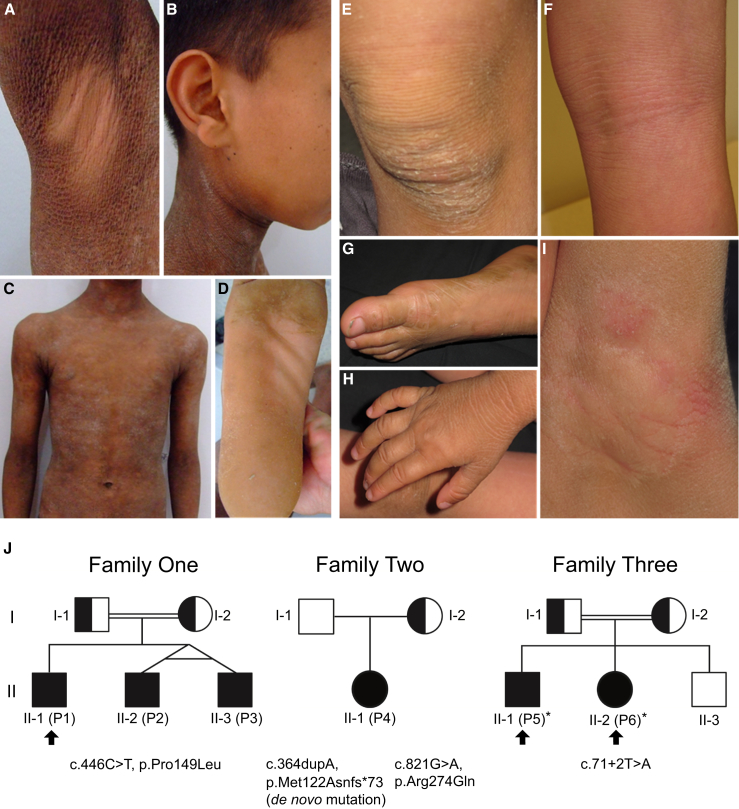
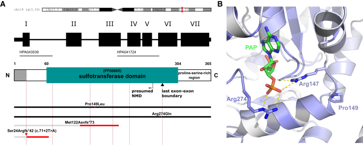
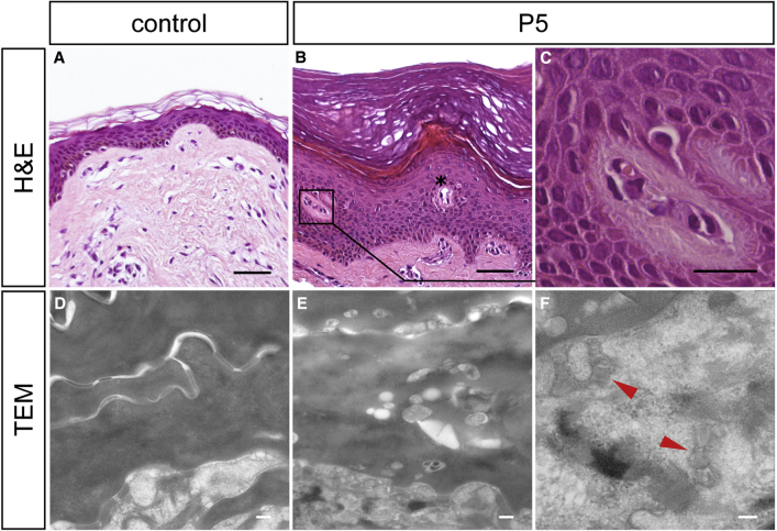
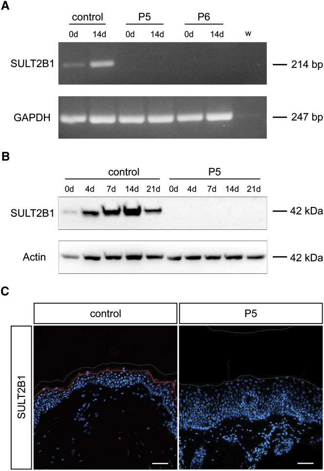
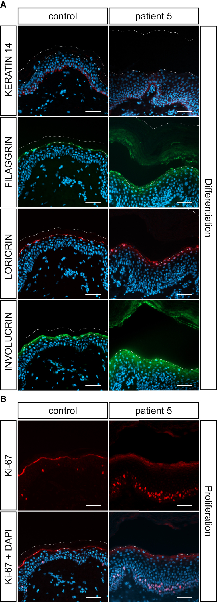
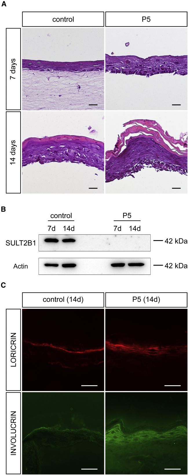
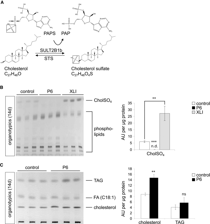
Ichthyoses are a clinically and genetically heterogeneous group of genodermatoses associated with abnormal scaling of the skin over the whole body. Mutations in nine genes are known to cause non-syndromic forms of autosomal-recessive congenital ichthyosis (ARCI). However, not all genetic causes for ARCI have been discovered to date. Using whole-exome sequencing (WES) and multigene panel screening, we identified 6 ARCI-affected individuals from three unrelated families with mutations in Sulfotransferase family 2B member 1 (SULT2B1), showing their causative association with ARCI. Cytosolic sulfotransferases form a large family of enzymes that are involved in the synthesis and metabolism of several steroids in humans. We identified four distinct mutations including missense, nonsense, and splice site mutations. We demonstrated the loss of SULT2B1 expression at RNA and protein levels in keratinocytes from individuals with ARCI by functional analyses. Furthermore, we succeeded in reconstructing the morphologic skin alterations in a 3D organotypic tissue culture model with SULT2B1-deficient keratinocytes and fibroblasts. By thin layer chromatography (TLC) of extracts from these organotypic cultures, we could show the absence of cholesterol sulfate, the metabolite of SULT2B1, and an increased level of cholesterol, indicating a disturbed cholesterol metabolism of the skin upon loss-of-function mutation in SULT2B1. In conclusion, our study reveals an essential role for SULT2B1 in the proper development of healthy human skin. Mutation in SULT2B1 leads to an ARCI phenotype via increased proliferation of human keratinocytes, thickening of epithelial layers, and altered epidermal cholesterol metabolism.

Keywords: 3D skin tissue culture model; SULT2B1; autosomal-recessive congenital ichthyosis; cholesterol sulfate cycle; epidermal differentiation; epidermal proliferation; epidermis; exome sequencing; ichthyosis; skin permeability barrier.


Figures

Figure 1
Figure 1
Clinical Features, Pedigrees, and SULT2B1 Mutations of Individuals with ARCI (A–D) At the age of 15 years, individual P1 (II-1, family 1) presents lamellar ichthyosis with generalized scaling and large brownish to gray scales. Hyperkeratotic plaques over the trunk (C) varied from mild to severe grade. A few areas such as the axilla (A), the face and ears (B), and the middle part of the soles (D) were not affected. (E–I) Individuals P5 (II-1, family 3) at the age of 10 years and P6 (II-2, family 3) at the age of 6 years also show sparing of popliteal fossa (P5 shown in F) and axilla region (P6 shown in I) with a milder phenotype than individual P1. The axillary region appeared macerated. The back of feet (P5 shown in G) and hands (P6 shown in H) and knees (P6 shown in E) showed very dry skin and were more severely affected. Lichenification of the popliteal fossa in individual P5 shown in (F). (J) Pedigrees of the three families and SULT2B1 mutations. In family 1, the homozygous missense mutation c.446C>T (p.Pro149Leu) was identified in individuals P1, P2, and P3, transmitted by consanguineous parents. In family 2, the only affected descendant, individual P4 (II-1), from non-consanguineous parents is compound heterozygous for the maternal inherited missense mutation c.821G>A (p.Arg274Gln) and a de novo insertion mutation with frameshift introducing a premature stop codon c.364dupA (p.Met122Asnfs73). Individuals P5 and P6 of family 3 are homozygous for the splice site mutation c.71+2T>A (p.Ser24Argfs42), which was transmitted by consanguineous parents. Clinical pictures from individuals P1, P5, and P6 are shown (arrows). Skin biopsies were obtained from individuals P5 and P6 (asterisks).
Figure 2
Figure 2
Schematic Representation of SULT2B1 Structure, Positions of ARCI-Causing Mutations, and Localization of the Missense Mutations in the PAPS/PAP Binding Pocket of SULT2B1 in the Wild-Type Crystal Structure (A) Localization of SULT2B1 on chromosome 19q13.33, exon/intron organization in SULT2B1, domain structures in SULT2B1, and positions of mutations. Dashed lines indicate exon/intron boundaries (rose), black bars fully expressed mutated proteins, and gray bars truncated proteins whose expression is assumed to be suppressed by NMD or other mechanisms. A proline-rich hydrophobic region (aa 305–365) is indicated at the C terminus of the protein. Near the C terminus, a black inverted triangle marks the position of the last exon-exon boundary. A dashed arrow labeled “NMD” (nonsense-mediated mRNA decay) indicates the ∼55 bp (∼18 aa) position upstream of the last exon-exon boundary. Assuming that the mutation c.71+2T>A (red triangle) leads to intron retention of the first intron, we have marked the hypothetical protein sequence leading to p.Ser24Argfs42. The epitopes of two SULT2B1 antibodies used in this study (HPA043539 and HPA041724, located at the N terminus and in the sulfotransferase domain, respectively) are indicated as black solid lines. (B) The characteristic proline side chain at position 149 (Pro149) stabilizes a bending of the protein backbone, which places the positively charged arginine at position 147 (Arg147, blue) in the proper position for a strong polar interaction with the negatively charged 3′ phosphate group of PAPS/PAP (PAPS/PAP in green-blue, phosphate group in red-orange, polar interactions as yellow dashed lines). Arg274 (blue) interacts strongly with the same phosphate group of PAPS. The intensity of blue coloring of the protein backbone reflects the conservation grade of the amino acid residues. The darker the blue color of the backbone, the more conserved the amino acid residue. Amino acid residues no. 276–282 and 306–311 were hidden for visualization purposes.
Figure 3
Figure 3
Histological and Ultrastructural Analysis of Skin Biopsies (A–C) Hematoxylin and eosin (H&E) staining of FFPE sections from (A) a healthy individual and (B, C) individual P5. Note the pronounced hyperkeratosis with a massive orthokeratotic cornified layer and a prominent granular layer in the epidermis of individual P5 (B). Blood vessels extended to epidermal layers (asterisk), frequently accompanied by (C) perivascular lymphocytic infiltrations with eosinophilic granulocytes appearing in the stratum spinosum and at the stratum spinosum-stratum granulosum junction. (D–F) Transmission electron microscopy (TEM). (D) Horny lamellae of healthy skin showing amorphous horny material of homogeneous electron density. (E) Horny lamellae of individual P6 showing variable electron densities of horny material and numerous small vesicular inclusions, putatively representing irregularly processed lamellar body contents in the horny lamellae. (F) Lamellated structures (arrowheads) within lamellar body profiles in skin of individual P6. Scale bars represent 50 μm in (A) and (B), 10 μm in (C), 200 nm in (D) and (E), and 100 nm in (F).
Figure 4
Figure 4
SULT2B1 mRNA and Protein Expression in Affected Individuals Is Disturbed (A) Absence of SULT2B1 expression at the mRNA level shown by RT-PCR on total RNA isolated from control cell line NHEK and keratinocytes from individuals P5 and P6 before differentiation (0d) and upon differentiation (14d). GAPDH served as positive control gene. Both samples of affected individuals did not result in a product for SULT2B1 whereas the control produced a weak signal at day 0 and an intense signal at day 14 of differentiation. (B) Western blot analysis of SULT2B1 in control keratinocytes and those of individual P5 upon in vitro differentiation (day 0, 4, 7, 14, 21) using an antibody raised against the sulfotransferase domain of the protein. Actin served as loading control. In control keratinocytes SULT2B1 was detected at 42 kDa with increasing expression during the differentiation process until a maximum at day 14. In keratinocytes of individual P5, SULT2B1 was not detectable. (C) Absence of SULT2B1 expression in the epidermis of individual P5 shown in vivo by immunofluorescence staining of FFPE sections. The healthy control epidermis shows strong expression of SULT2B1 in the granular layer (red). Counterstaining with DAPI (blue); dashed lines indicate the stratum granulosum-stratum corneum junction as well as the upper border of the stratum corneum. Abbreviations are as follows: w, water control; kDa, kilodalton; DAPI, 4’,6-diamidino-2-phenylindole. Scale bars represent 50 μm.
Figure 5
Figure 5
Disturbed Epidermal Proliferation and Differentiation upon SULT2B1 Depletion Shown by Immunofluorescence Staining with Antibodies Specific for Keratin 14, Filaggrin, Loricrin, Involucrin, and Ki-67 (A) FFPE sections from individual P5 show increased expression of terminal differentiation markers filaggrin, loricrin, and the early differentiation marker involucrin compared to control skin. Expression of the marker for undifferentiated keratinocytes, keratin 14, appears normal in the epidermis of individual P5. The stratum corneum in the individual’s skin is massively thickened, but often detached during staining procedure. (B) Staining of FFPE sections with a nuclear marker for proliferation, Ki-67, indicates strongly enhanced proliferation in the epidermal basal layer of individual P5 in comparison to control skin. Counterstaining with DAPI (blue); dashed lines indicate the stratum granulosum-stratum corneum junction as well as the upper border of the stratum corneum. Scale bars represent 50 μm.
Figure 6
Figure 6
Reconstitution of Affected Individual’s Skin Morphology in a 3D Organotypic Tissue Culture Model (A) Hematoxylin and eosin staining of FFPE sections from the organotypic model of individual P5 reflects the thickening of the stratum corneum and the prominent granular layer compared to control 14 days after induction of differentiation. The dermal component was often detached during staining procedure (except the control at 7 days). (B) Western blot analysis of SULT2B1 with organotypic models from control and mutated cell line confirms the absence of SULT2B1 in individual P5. Actin served as loading control. (C) Immunofluorescence staining of cryo sections from organotypic cultures upon differentiation (14 days) verified increased expression of differentiation markers loricrin and involucrin in the model of individual P5. Abbreviation: kDa, kilodalton. Scale bars represent 100 μm in (A) and 50 μm in (C).
Figure 7
Figure 7
Mutated SULT2B1 Causes Imbalance in Cholesterol Metabolism (A) Enzymatic conversion of unconjugated cholesterol to cholesterol sulfate (drawn from Chem3D 15.1, PerkinElmer). SULT2B1 catalyzes the transfer of the 5′ sulfate from the donor 3′-phosphoadenosine 5′-phosphosulfate (PAPS) to the hydroxyl group (OH) of cholesterol; consequently, cholesterol is converted to cholesterol sulfate by sulfonation. STS catalyzes the counter reaction converting cholesterol sulfate into cholesterol. (B) TLC of polar lipids (left) and statistical analysis (right) of organotypic extracts from individual P6, the XLI-affected person, and control organotypic extracts upon differentiation (14 days) revealed a statistically significant increased amount of cholesterol sulfate in the XLI-affected person and its complete absence in individual P6. Lipids were normalized to corresponding 400 μg of cellular protein. (C) TLC of neutral lipids (left) and statistical analysis (right) of extracts from individual P6 and control organotypic models upon differentiation (14 days) resulted in a statistically significant increased amount of cholesterol in individual P6. Lipids were normalized to corresponding 100 μg of cellular protein. Data are presented as means + SD of triplicates and are representative for three independent experiments. Statistical significance was examined by unpaired two-tailed Student’s t test (∗∗p < 0.01; ∗∗∗p < 0.001; ns, p > 0.05). Abbreviations are as follows: CholSO4, cholesterol sulfate; TAG, triacylglycerol; FA, fatty acid (C18:1) Oleate cis-9-Octadecenoate; n.d., not detectable; ns, not significant.

References

    1. Oji V., Tadini G., Akiyama M., Blanchet Bardon C., Bodemer C., Bourrat E., Coudiere P., DiGiovanna J.J., Elias P., Fischer J. Revised nomenclature and classification of inherited ichthyoses: results of the First Ichthyosis Consensus Conference in Sorèze 2009. J. Am. Acad. Dermatol. 2010;63:607–641. - PubMed
    1. Traupe H., Fischer J., Oji V. Nonsyndromic types of ichthyoses - an update. J. Dtsch. Dermatol. Ges. 2014;12:109–121. - PubMed
    1. Huber M., Rettler I., Bernasconi K., Frenk E., Lavrijsen S.P., Ponec M., Bon A., Lautenschlager S., Schorderet D.F., Hohl D. Mutations of keratinocyte transglutaminase in lamellar ichthyosis. Science. 1995;267:525–528. - PubMed
    1. Russell L.J., DiGiovanna J.J., Rogers G.R., Steinert P.M., Hashem N., Compton J.G., Bale S.J. Mutations in the gene for transglutaminase 1 in autosomal recessive lamellar ichthyosis. Nat. Genet. 1995;9:279–283. - PubMed
    1. Jobard F., Lefèvre C., Karaduman A., Blanchet-Bardon C., Emre S., Weissenbach J., Ozgüc M., Lathrop M., Prud’homme J.-F., Fischer J. Lipoxygenase-3 (ALOXE3) and 12(R)-lipoxygenase (ALOX12B) are mutated in non-bullous congenital ichthyosiform erythroderma (NCIE) linked to chromosome 17p13.1. Hum. Mol. Genet. 2002;11:107–113. - PubMed

MeSH terms